Electroporation
Electroporation, or electropermeabilization, is a microbiology technique in which an electrical field is applied to cells in order to increase the permeability of the cell membrane, allowing chemicals, drugs, or DNA to be introduced into the cell.[1] In microbiology, the process of electroporation is often used to transform bacteria, yeast, or plant protoplasts by introducing new coding DNA. If bacteria and plasmids are mixed together, the plasmids can be transferred into the bacteria after electroporation, though depending on what is being transferred cell-penetrating peptides or CellSqueeze could also be used. Electroporation works by passing thousands of volts across a distance of one to two millimeters of suspended cells in an electroporation cuvette (1.0 – 1.5 kV, 250 – 750V/cm). Afterwards, the cells have to be handled carefully until they have had a chance to divide, producing new cells that contain reproduced plasmids. This process is approximately ten times more effective than chemical transformation.[1][2]
Electroporation is also highly efficient for the introduction of foreign genes into tissue culture cells, especially mammalian cells. For example, it is used in the process of producing knockout mice, as well as in tumor treatment, gene therapy, and cell-based therapy. The process of introducing foreign DNA into eukaryotic cells is known as transfection. Electroporation is highly effective for transfecting cells in suspension using electroporation cuvettes. Electroporation has proven efficient for use on tissues in vivo, for in utero applications as well as in ovo transfection. Adherent cells can also be transfected using electroporation, providing researchers with an alternative to trypsinizing their cells prior to transfection. One downside to electroporation, however, is that after the process the gene expression of over 7,000 genes can be affected.[3] This can cause problems in studies where gene expression has to be controlled to ensure accurate and precise results.
Cell fusion is of interest not only as an essential process in cell biology, but also as a useful method in biotechnology and medicine. Artificially induced fusion can be used to investigate and treat different diseases, like diabetes[4][5][6] regenerate axons of the central nerve system,[7] and produce cells with desired properties, such as in cell vaccines for cancer immunotherapy.[8][9][10] However, the first and most known application of cell fusion is production of monoclonal antibodies in hybridoma technology, where hybrid cell lines (hybridomas) are formed by fusing specific antibody-producing B lymphocytes with a myeloma (B lymphocyte cancer) cell line.[11][12]
Laboratory practice

Electroporation is performed with electroporators, purpose-built appliances which create an electrostatic field in a cell solution. The cell suspension is pipetted into a glass or plastic cuvette which has two aluminium electrodes on its sides. For bacterial electroporation, typically a suspension of around 50 microliters is used. Prior to electroporation, this suspension of bacteria is mixed with the plasmid to be transformed. The mixture is pipetted into the cuvette, the voltage and capacitance are set, and the cuvette is inserted into the electroporator. The process requires direct contact between the electrodes and the suspension. Immediately after electroporation, one milliliter of liquid medium is added to the bacteria (in the cuvette or in an Eppendorf tube), and the tube is incubated at the bacteria's optimal temperature for an hour or more to allow recovery of the cells and expression of the plasmid, followed by bacterial culture on agar plates.
The success of the electroporation depends greatly on the purity of the plasmid solution, especially on its salt content. Solutions with high salt concentrations might cause an electrical discharge (known as arcing), which often reduces the viability of the bacteria. For a further detailed investigation of the process, more attention should be paid to the output impedance of the porator device and the input impedance of the cells suspension (e.g. salt content).
Since the cell membrane is not able to pass current (except in ion channels), it acts as an electrical capacitor. Subjecting membranes to a high-voltage electric field results in their temporary breakdown, resulting in pores that are large enough to allow macromolecules (such as DNA) to enter or leave the cell.[13]
Additionally, electroporation can be used to increase permeability of cells during in Utero injections and surgeries. Particularly, the electroporation allows for a more efficient transfection of DNA, RNA, shRNA, and all nucleic acids into the cells of mice and rats. The success of in vivo electroporation depends greatly on voltage, repetition, pulses, and duration. Developing central nervous systems are most effective for in vivo electroporation due to the visibility of ventricles for injections of nucleic acids, as well as the increased permeability of dividing cells. Electroporation of injected in utero embryos is performed through the uterus wall, often with forceps-type electrodes to limit damage to the embryo. [14]
Medical applications
The first group to look at electroporation for medical applications was led by Lluis M Mir at the Institute Gustave Roussy. In this case, they looked at the use of reversible electroporation in conjunction with impermeable macromolecules. The first research looking at how nanosecond pulses might be used on human cells was conducted by researchers at Eastern Virginia Medical School and Old Dominion University, and published in 2003.[15]
With regards to irreversible electroporation, the first successful treatment of malignant cutaneous tumors implanted in mice was completed in 2007 by a group of scientists who achieved complete tumor ablation in 12 out of 13 mice. They accomplished this by sending 80 pulses of 100 microseconds at 0.3 Hz with an electrical field magnitude of 2500 V/cm to treat the cutaneous tumors.[16]
A higher voltage of electroporation was found in pigs to irreversibly destroy target cells within a narrow range while leaving neighboring cells unaffected, and thus represents a promising new treatment for cancer, heart disease and other disease states that require removal of tissue.[17] Irreversible electroporation (IRE) has since proven effective in treating human cancer, with surgeons at Johns Hopkins and other institutions now using the technology to treat pancreatic cancer previously thought to be unresectable.[18]
A recent technique called non-thermal irreversible electroporation (N-TIRE) has proven successful in treating many different types of tumors and other unwanted tissue. This procedure is done using small electrodes (about 1mm in diameter), placed either inside or surrounding the target tissue to apply short, repetitive bursts of electricity at a predetermined voltage and frequency. These bursts of electricity increase the resting transmembrane potential (TMP), so that nanopores form in the plasma membrane. When the electricity applied to the tissue is above the electric field threshold of the target tissue, the cells become permanently permeable from the formation of nanopores. As a result, the cells are unable to repair the damage and die due to a loss of homeostasis.[19] N-TIRE is unique to other tumor ablation techniques in that it does not create thermal damage to the tissue around it.
Contrastingly, reversible electroporation occurs when the electricity applied with the electrodes is below the electric field threshold of the target tissue. Because the electricity applied is below the cells' threshold, it allows the cells to repair their phospholipid bilayer and continue on with their normal cell functions. Reversible electroporation is typically done with treatments that involve getting a drug or gene (or other molecule that is not normally permeable to the cell membrane) into the cell. Not all tissue has the same electric field threshold; therefore careful calculations need to be made prior to a treatment to ensure safety and efficacy.[20]
One major advantage of using N-TIRE is that, when done correctly according to careful calculations, it only affects the target tissue. Proteins, the extracellular matrix, and critical structures such as blood vessels and nerves are all unaffected and left healthy by this treatment. This allows for a quicker recovery, and facilitates a more rapid replacement of dead tumor cells with healthy cells.[21]
Before doing the procedure, scientists must carefully calculate exactly what needs to be done, and treat each patient on an individual case-by-case basis. To do this, imaging technology such as CT scans and MRI's are commonly used to create a 3D image of the tumor. From this information, they can approximate the volume of the tumor and decide on the best course of action including the insertion site of electrodes, the angle they are inserted in, the voltage needed, and more, using software technology. Often, a CT machine will be used to help with the placement of electrodes during the procedure, particularly when the electrodes are being used to treat tumors in the brain.[22]
The entire procedure is very quick, typically taking about five minutes. The success rate of these procedures is high and is very promising for future treatment in humans. One disadvantage to using N-TIRE is that the electricity delivered from the electrodes can stimulate muscle cells to contract, which could have lethal consequences depending on the situation. Therefore, a paralytic agent must be used when performing the procedure. The paralytic agents that have been used in such research are successful; however, there is always some risk, albeit slight, when using anesthetics.
A more recent technique has been developed called high-frequency irreversible electroporation (H-FIRE). This technique uses electrodes to apply bipolar bursts of electricity at a high frequency, as opposed to unipolar bursts of electricity at a low frequency. This type of procedure has the same tumor ablation success as N-TIRE. However, it has one distinct advantage, H-FIRE does not cause muscle contraction in the patient and therefore there is no need for a paralytic agent.[23]
Drug and gene delivery
Electroporation can also be used to help deliver drugs or genes into the cell by applying short and intense electric pulses that transiently permeabilize cell membrane, thus allowing transport of molecules otherwise not transported through a cellular membrane. This procedure is referred to as electrochemotherapy when the molecules to be transported are chemotherapeutic agents or gene electrotransfer when the molecule to be transported is DNA. Scientists from Karolinska Institutet and the University of Oxford use electroporation of exosomes to deliver siRNAs, antisense oligonucleotides, chemotherapeutic agents and proteins specifically to neurons after inject them systemically (in blood). Because these exosomes are able to cross the blood brain barrier, this protocol could solve the problem of poor delivery of medications to the central nervous system, and potentially treat Alzheimer's, Parkinson's Disease and brain cancer, among other conditions.[24]
Physical mechanism
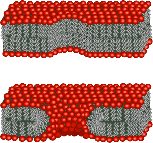
Electroporation allows cellular introduction of large highly charged molecules such as DNA which would never passively diffuse across the hydrophobic bilayer core.[1] This phenomenon indicates that the mechanism is the creation of nm-scale water-filled holes in the membrane. Although electroporation and dielectric breakdown both result from application of an electric field, the mechanisms involved are fundamentally different. In dielectric breakdown the barrier material is ionized, creating a conductive pathway. The material alteration is thus chemical in nature. In contrast, during electroporation the lipid molecules are not chemically altered but simply shift position, opening up a pore which acts as the conductive pathway through the bilayer as it is filled with water.
Electroporation is a dynamic phenomenon that depends on the local transmembrane voltage at each point on the cell membrane. It is generally accepted that for a given pulse duration and shape, a specific transmembrane voltage threshold exists for the manifestation of the electroporation phenomenon (from 0.5 V to 1 V). This leads to the definition of an electric field magnitude threshold for electroporation (Eth). That is, only the cells within areas where E≧Eth are electroporated. If a second threshold (Eir) is reached or surpassed, electroporation will compromise the viability of the cells, i.e., irreversible electroporation (IRE).[25]
Electroporation is a multi-step process with several distinct phases.[26] First, a short electrical pulse must be applied. Typical parameters would be 300–400 mV for < 1 ms across the membrane (note- the voltages used in cell experiments are typically much larger because they are being applied across large distances to the bulk solution so the resulting field across the actual membrane is only a small fraction of the applied bias). Upon application of this potential the membrane charges like a capacitor through the migration of ions from the surrounding solution. Once the critical field is achieved there is a rapid localized rearrangement in lipid morphology. The resulting structure is believed to be a "pre-pore" since it is not electrically conductive but leads rapidly to the creation of a conductive pore.[27] Evidence for the existence of such pre-pores comes mostly from the "flickering" of pores, which suggests a transition between conductive and insulating states.[28] It has been suggested that these pre-pores are small (~3 Å) hydrophobic defects. If this theory is correct, then the transition to a conductive state could be explained by a rearrangement at the pore edge, in which the lipid heads fold over to create a hydrophilic interface. Finally, these conductive pores can either heal, resealing the bilayer or expand, eventually rupturing it. The resultant fate depends on whether the critical defect size was exceeded[29] which in turn depends on the applied field, local mechanical stress and bilayer edge energy.
References
- 1 2 3 Neumann, E; Schaefer-Ridder, M; Wang, Y; Hofschneider, PH (1982). "Gene transfer into mouse lyoma cells by electroporation in high electric fields". The EMBO Journal. 1 (7): 841–5. PMC 553119. PMID 6329708.
- ↑ Sugar, I.P.; Neumann, E. (1984). "Stochastic model for electric field-induced membrane pores electroporation". Biophysical Chemistry. 19 (3): 211–25. doi:10.1016/0301-4622(84)87003-9. PMID 6722274.
- ↑ Anne Trafton (2 February 2016). "Cell squeezing enhances protein imaging". MIT News Office.
- ↑ McClenaghan, N. H. Physiological regulation of the pancreatic β-cell: functional insights for understanding and therapy of diabetes. Exp. Physiol. 92, 481–496 (2007).
- ↑ Yanai, G. et al. Electrofusion of mesenchymal stem cells and islet cells for diabetes therapy: A rat model. PLoS ONE 8, e64499 (2013).
- ↑ McCluskey, J. T. et al. Development and functional characterization of insulin-releasing human pancreatic beta cell lines produced by electrofusion. J. Biol. Chem. 286, 21982–21992 (2011).
- ↑ Sretavan, D. W., Chang, W., Hawkes, E., Keller, C. & Kliot, M. Microscale surgery on single axons. Neurosurgery 57, 635–646 (2005).
- ↑ Guo, W., Guo, Y., Tang, S., Qu, H. & Zhao, H. Dendritic cell-Ewing's sarcoma cell hybrids enhance antitumor immunity. Clin. Orthop. 466, 2176–2183 (2008).
- ↑ Koido, S. et al. Regulation of tumor immunity by tumor/dendritic cell fusions. Clin. Dev. Immunol. 2010, 516768 (2010).
- ↑ Avigan, D., Rosenblatt, J. & Kufe, D. Dendritic/tumor fusion cells as cancer vaccines. Semin. Oncol. 39, 287–295 (2012).
- ↑ Vor dem Esche, U. et al. Passive vaccination with a human monoclonal antibody: generation of antibodies and studies for efficacy in Bacillus anthracis infections. Immunobiology 216, 847–853 (2011).
- ↑ Trontelj, K. et al. Optimization of bulk cell electrofusion in vitro for production of human-mouse heterohybridoma cells. Bioelectrochemistry 74, 124–129 (2008).
- ↑ Potter, Huntington; Heller, Richard (2003-05-01). "Transfection by Electroporation". Current Protocols in Molecular Biology. CHAPTER: Unit–9.3. doi:10.1002/0471142727.mb0903s62. ISSN 1934-3639. PMC 2975437. PMID 18265334.
- ↑ "Embryonic In Vivo Electroporation in the Mouse". Methods in Enzymology. 477: 37–50. 2010-01-01. doi:10.1016/S0076-6879(10)77003-8. ISSN 0076-6879.
- ↑ Beebe SJ, Fox PM, Rec LJ, Willis EL, Schoenbach KH (August 2003). "Nanosecond, high-intensity pulsed electric fields induce apoptosis in human cells". FASEB J. 17 (11): 1493–5. doi:10.1096/fj.02-0859fje. PMID 12824299.
- ↑ Al-Sakere, Bassim; André, Franck; Bernat, Claire; Connault, Elisabeth; Opolon, Paule; Davalos, Rafael V.; Rubinsky, Boris; Mir, Lluis M. (2007). Isalan, Mark, ed. "Tumor Ablation with Irreversible Electroporation". PLoS ONE. 2 (11): e1135. Bibcode:2007PLoSO...2.1135A. doi:10.1371/journal.pone.0001135. PMC 2065844. PMID 17989772.
- ↑ Sarah Yang (2007-02-12). "New medical technique punches holes in cells, could treat tumors". Retrieved 2007-12-13.
- ↑ "A Potential Boon for Pancreatic Cancer Patients". Johns Hopkins Surgery: News From the Johns Hopkins Department of Surgery. 2014-06-23.
- ↑ Garcia, Paulo A.; Rossmeisl, John H.; Davalos, Rafael V. (2011). "Electrical conductivity changes during irreversible electroporation treatment of brain cancer". 2011 Annual International Conference of the IEEE Engineering in Medicine and Biology Society: 739–42. doi:10.1109/IEMBS.2011.6090168. ISBN 978-1-4577-1589-1. PMID 22254416.
- ↑ Garcia, P A; Neal, Robert E; Rossmeisl, John H; Davalos, R V (2010). "Non-thermal irreversible electroporation for deep intracranial disorders". 2010 Annual International Conference of the IEEE Engineering in Medicine and Biology: 2743–6. doi:10.1109/IEMBS.2010.5626371. ISBN 978-1-4244-4123-5. PMID 21095962.
- ↑ Garcia, Paulo A.; Rossmeisl, John H.; Neal, Robert E.; Ellis, Thomas L.; Olson, John D.; Henao-Guerrero, Natalia; Robertson, John; Davalos, Rafael V. (2010). "Intracranial Nonthermal Irreversible Electroporation: In Vivo Analysis". The Journal of Membrane Biology. 236 (1): 127–36. doi:10.1007/s00232-010-9284-z. PMID 20668843.
- ↑ Neal, R E; Garcia, P A; Rossmeisl, J H; Davalos, R V (2010). "A study using irreversible electroporation to treat large, irregular tumors in a canine patient". 2010 Annual International Conference of the IEEE Engineering in Medicine and Biology: 2747–50. doi:10.1109/IEMBS.2010.5626372. ISBN 978-1-4244-4123-5. PMID 21095963.
- ↑ Arena, Christopher B; Sano, Michael B; Rossmeisl, John H; Caldwell, John L; Garcia, Paulo A; Rylander, Marissa; Davalos, Rafael V (2011). "High-frequency irreversible electroporation (H-FIRE) for non-thermal ablation without muscle contraction". BioMedical Engineering OnLine. 10: 102. doi:10.1186/1475-925X-10-102. PMC 3258292. PMID 22104372.
- ↑ El-Andaloussi S, Lee Y, Lakhal-Littleton S, Li J, Seow Y, Gardiner C, Alvarez-Erviti L, Sargent IL, Wood MJ (December 2012). "Exosome-mediated delivery of siRNA in vitro and in vivo". Nat Protoc. 7 (12): 2112–26. doi:10.1038/nprot.2012.131. PMID 23154783.
- ↑ Ivorra, Antoni; Rubinsky, Boris. "Gels with predetermined conductivity used in electroporation of tissue USPTO Application #: 20080214986 — Class: 604 21 (USPTO)".
- ↑ Weaver, James C.; Chizmadzhev, Yu.A. (1996). "Theory of electroporation: A review". Bioelectrochemistry and Bioenergetics. 41 (2): 135–60. doi:10.1016/S0302-4598(96)05062-3.
- ↑ Becker, S. M.; Kuznetsov, A. V. (2007). "Local Temperature Rises Influence in Vivo Electroporation Pore Development: A Numerical Stratum Corneum Lipid Phase Transition Model". Journal of Biomechanical Engineering. 129 (5): 712–21. doi:10.1115/1.2768380. PMID 17887897.
- ↑ Melikov, Kamran C.; Frolov, Vadim A.; Shcherbakov, Arseniy; Samsonov, Andrey V.; Chizmadzhev, Yury A.; Chernomordik, Leonid V. (2001). "Voltage-Induced Nonconductive Pre-Pores and Metastable Single Pores in Unmodified Planar Lipid Bilayer". Biophysical Journal. 80 (4): 1829–36. Bibcode:2001BpJ....80.1829M. doi:10.1016/S0006-3495(01)76153-X. PMC 1301372. PMID 11259296.
- ↑ Joshi, R.; Schoenbach, K. (2000). "Electroporation dynamics in biological cells subjected to ultrafast electrical pulses: A numerical simulation study". Physical Review E. 62 (1 Pt B): 1025–33. Bibcode:2000PhRvE..62.1025J. doi:10.1103/PhysRevE.62.1025. PMID 11088559.