Dextrocardia
| Dextrocardia | |
|---|---|
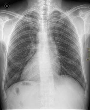 | |
| Chest X ray of a person with dextrocardia situs inversus showing the cardiac apex facing the right | |
| Specialty |
Medical genetics |
Dextrocardia (from Latin dexter, meaning "right," and Greek kardia, meaning "heart") is a rare congenital condition in which the apex of the heart is located on the right side of the body.[1] There are two main types of dextrocardia: dextrocardia of embryonic arrest (also known as isolated dextrocardia) and dextrocardia situs inversus. Dextrocardia situs inversus is further divided.
Classification
Dextrocardia of embryonic arrest
In this form of dextrocardia, the heart is simply placed further right in the thorax than is normal. It is commonly associated with severe defects of the heart and related abnormalities including pulmonary hypoplasia.[2]
Dextrocardia situs inversus
Dextrocardia with situs inversus refers to the heart being a mirror image situated on the right side. For all visceral organs to be mirrored, the correct term is dextrocardia situs inversus totalis.
Although statistically people with dextrocardia do not have any medical problems from the disorder, they may be prone to a number of bowel, esophageal, bronchial and cardiovascular disorders (such as double outlet right ventricle, endocardial cushion defect and pulmonary stenosis).[3] Certain cardiovascular and pulmonary disorders related to dextrocardia can be life-threatening if left unchecked (see reference).
Kartagener syndrome may also be present in patients with dextrocardia but this must be in the setting of situs inversus and may include male infertility.[4]
Diagnosis
Medical diagnosis of the two forms of congenital dextrocardia can be made by ECG[2] or imaging.
Technical dextrocardia
Technical dextrocardia refers to an ECG reading that has no basis in the patient's anatomy. This apparent presentation is typically caused by the accidental lead placement of the left and right arm electrodes. Usually this would show as an extreme axis deviation.
Management
ECG leads must be placed in reversed positions on a person with dextrocardia. In addition, when defibrillating someone with dextrocardia, the pads should be placed in reverse positions.[5] That is, instead of upper right and lower left, pads should be placed upper left and lower right.[6]
When heart transplantation is required in a person with situs inversus, reconstruction of the venous pathways to accommodate a normal donor heart is a major, but not insurmountable, challenge.[7]
Epidemiology
Dextrocardia is believed to occur in approximately 1 in 12,019 pregnancies.[8]
A Japanese study of 1,753 fetal cardiac echocardiograms over five years only revealed two cases.[9]
References
- ↑ Stephenson, Susan (2012). Diagnostic Medical Sonography Obstetrics and Gynecology. 351 West Camden Street Baltimore, MD 21201: Lippincott Williams & Wilkins. p. 582.
- 1 2 M. E. Abbott & J. C. Meakins (1915). "On the differentiation of two forms of congenital dextrocardia". Bulletin of the International Association of Medical Museums (5): 134–138.
- ↑ MedlinePlus Medical Dictionary. "Dextrocardia".
- ↑ Renee A, Laux MS. "Kartagener Syndrome".
- ↑ Bindra, S. MD; Tabibiazar, R. MD; Mazar, M MD & Dave, R MD (2011). "Clinical Vignette: Dextrocardia with Situs Inversus: Through the Looking Glass with an ECG". Proceedings of UCLA Healthcare. Department of Medicine, UCLA. 15.
- ↑ Eddy, S. "Dextrocardia and Proper Lead Placement".
- ↑ Deuse, Tobias; Reitz, Bruce A. (September 2009). "Heart-Lung Transplantation In Situs Inversus Totalis". The Annals of Thoracic Surgery. 88 (3): 1002–1003. doi:10.1016/j.athoracsur.2009.01.060. PMID 19699943.
- ↑ Bohun CM, Potts JE, Casey BM, Sandor GG (July 2007). "A population-based study of cardiac malformations and outcomes associated with dextrocardia". Am. J. Cardiol. 100 (2): 305–9. doi:10.1016/j.amjcard.2007.02.095. PMID 17631088.
- ↑ Sato, T.; et al. (2014). "Clinical Course and Prognosis of Minor Abnormal Sonographic Findings in Fetal Echocardiography: Five Years of Experience at a Single Institute". Pediatric Cardiology and Cardiac Surgery (in Japanese and English). 30 (5): 563–568. doi:10.9794/jspccs.30.563.
External links
| Classification | |
|---|---|
| External resources |
- Dextrocardia at NIH's Office of Rare Diseases
- Dextrocardia with situs inversus at NIH's Office of Rare Diseases
| Side | Left | Both | Right |
|---|---|---|---|
| General | Ambidexterity | ||
| In cognitive abilities | Geschwind–Galaburda hypothesis | ||
| In brain | |||
| In eyes | Ocular dominance | ||
| In hands | Left-handedness | Cross-dominance | Right-handedness |
| Handedness in boxing | Southpaw stance | Orthodox stance | |
| Handedness in people | Musicians | ||
| Handedness related to | |||
| Handedness measurement | Edinburgh Handedness Inventory | ||
| Handedness genetics | LRRTM1 | ||
| In heart | Levocardia | Dextrocardia | |
| In major viscera | Situs solitus | Situs ambiguus | Situs inversus |
| In feet | Footedness | ||
| Footedness in surfing | Regular foot | Goofy foot | |