Dental anesthesia
| Dental anesthesia | |
|---|---|
| MeSH | D000766 |
Dental anesthesia (or dental anaesthesia) is a field of anesthesia that includes not only local anesthetics but sedation and general anesthesia.
Local anesthetic agents in dentistry
The most commonly used local anesthetic is lidocaine (also called xylocaine or lignocaine), a modern replacement for procaine (also known as novocaine). Its half-life in the body is about 1.5–2 hours. Other local anesthetic agents in current use include articaine (also called septocaine or ubistesin), bupivacaine (a long-acting anesthetic), Prilocaine (also called Citanest), and mepivacaine (also called Carbocaine or Polocaine). A combination of these may be used depending on the situation. Also, most agents come in two forms: with and without epinephrine (adrenaline) or other vasoconstrictor that allow the agent to last longer and also controls bleeding in the tissue during procedures. Usually the case is classified using the ASA Physical Status Classification System before any anesthesia is given.
Maxillary anaesthesia
Local anaesthesia is deposited at the buccal (cheek) side of the maxillary alveolus which can diffuse through the thin cortical plate of the maxilla, then further into the pulp of the tooth in order to achieve dental anaesthesia effect.
Buccal Infiltration
Instruments
- 27 or 30 gauge needle
- Short needle, usually 20-25mm long needle
- Syringe
- Local anaesthesia cartridge[1]
Technique
- Retract the lip/ cheek with a mirror or operator’s fingers. This is easier when the patient’s mouth is partially open as cheek is more relaxed, hence it’s easier to be stretched to gain access. A widely open mouth would result in over-stretching of the cheek across the teeth which limit the access.
- Identify the point of needle insertion.
- Use a gauze to clean the injection site and keep it dry before applying topical anaesthesia prior to injection.
 Maxillary infiltration
 - Ensure the lip/ cheek is stretched in a superior direction and the needle is then penetrated 45’ with the buccal cortical plate of the bone through the taut tissue of the muccobuccal fold.
- Direct the needle towards apex of the tooth.
- If the needle is in contact with the bone, withdraw the needle slightly before depositing the local anaesthesia.
- Aspiration should be carried out before injecting, this is to avoid intravascular injection. Injecting into vascular tissues could result in failed anaesthesia and increase risk of systemic side effects.
- The solution should be deposited slowly (1ml/min)
- Wait for at least 2 minutes before checking the site of anaesthesia with a dental probe.[1][2]
Limitations
- Effectiveness of an infiltration is determined by the permeability of the tissues through which the solution has to infiltrate and diffuse through. Thick cortical plate would reduce the distribution and diffusion of the solution through the bone.
- E.g. Thick zygomatic process adjacent to the upper first molar is one the most common causes of infiltration failure.
- Localised infection at the site of infiltration.
- Collateral supply of nasopalatine nerves/ greater palatine nerve to the pulp would affect the effectiveness of buccal infiltration.[1]
Advantages
- Straightforward and simple technique
- Reduce risk of intravascular injection
- All nerve endings in the area of local anaesthesia of deposition would be anaesthetised independent of the nerve origin if it’s successful.
- Sometimes it can be useful to achieve haemostasis if needed[1]
Disadvantages
- Pulpal anaesthesia can only be achieved and effective when the local anaesthetic is able to diffuse through the cortical bone.
- Infiltrating into a highly inflamed area may increase the risk of spread of local infection.
- Anaesthetic effect of the tissues is only restricted to the area of injection.[1]
Palatal infiltration
This is only needed prior to extraction or dental surgeries. Buccal infiltration would be sufficient to carry out most of the dental treatment.
Technique
- The operator should insert the needle from the opposite side of the patient.
- The point of injection should be 90’ to the palatine bone, in the fleshiest part of the palate around 1-1.5 cm from the gingival margin.
- Infiltrate 0.2-0.3ml of the solution slowly into the palatine mucosa just slightly distal of the tooth[1]
Disadvantage
- More painful/ discomfort in comparison to buccal infiltration.[3]
Regional block techniques
This anaesthesia technique that can be useful in the maxilla intra-orally when infiltration is not effective or multiple sites or large area of anaesthesia is required at a time. There are different types of regional block techniques specifically used in dentistry to achieve dental anaesthesia in required areas within the maxilla.[1]
- Posterior superior alveolar nerve block
- Maxillary molar nerve block
- Middle superior alveolar nerve block
- Anterior superior alveolar nerve block
- Infraorbital nerve block
- Palatal anterior superior alveolar nerve block
- Anterior middle superior alveolar nerve block
- Maxillary nerve block
- Greater palatine nerve block:[1][2]
- Anaesthetises the posterior 2/3 of soft tissues and bone of the hard palate from one side of the midline (maxillary 3rd molar to the canine region on the same side)
- Technique
- Locate the greater palatine foramen between the junction of the maxillary 2nd and 3rd molar.
- Apply topical anaesthesia.
- Point of needle insertion is adjacent to the greater palatine foramen, 90’ to the hard palate, only a few millimetres of the needle should be inserted.
- Aspirate before injecting the solution.
- Small amount of local anaesthetic is required, around 0.2mL is enough for the greater palatine nerve block.
- Nasopalatine nerve block:[1][2]
- Anaesthetises the anterior hard palate and the associated soft tissues in the incisors region bilaterally.
- Technique
- Ask patient to open the mouth widely.
- Locate the incisive foramen. The incisive papilla normally present as a slight raised of soft tissue, 5 mm posterior to the midline of upper central incisors, which overlies the incisive foramen.
- Apple topical anaesthesia.
- Insert needle 45’ to the palatal mucosa with the bevel facing the hard palate just lateral or one side into the crest of the incisive papilla. Penetrate the needle 3–4 mm into the incisive papilla or until the needle contacts bone.
- Aspirate before injecting the solution.
- Less than 0.2mL of local anaesthesia should be injected.
Mandibular anaesthesia
Both regional block and infiltration techniques are considered the first choice injections for anaesthetising the mandibular teeth.
Different techniques are chosen based on different factors:
- Patient age[1]
- Infiltration anaesthesia is a preferable method to anaesthetise deciduous/ primary teeth in children. The method is similar to the maxillary buccal infiltration. Ensure the lip/ cheek is stretched in a lateral and inferior direction instead of superiorly and the needle is then penetrated 45’ with the buccal cortical plate of the bone through the taut tissue of the muccobuccal fold.
- Tooth to be anaesthetised[1]
- Infiltration anaesthesia should be the first choice of method for pulpal and soft tissue anaesthesia of mandibular permanent incisors in adults. Regional block injections are sometimes ineffective due to crossover innervation from the opposite side of inferior alveolar nerve. It is recommended to deposit at least 0.5mL at each buccal and lingual site in the apical region of the tooth of interest. The use of infiltration anaesthesia with 4% articaine with 1:100,000 epinephrine in obtaining pulp anaesthesia of the mandibular permanent first molar is getting more common these days due to its successful formulation.
Regional block techniques
Inferior alveolar and lingual block
The inferior alveolar nerve block is probably one of the most common methods used by dentist to anaesthetise the mandibular teeth in adults. This technique aims to inject the needle and deposit local anaesthetic close the near to the nerve before it enters the mandibular foramen, which locates on the medial aspect of the mandibular ramus. This is to block the nerve transmission in the inferior alveolar nerve before entering into the bone through the mandibular foramen.[4]
Basic anatomy
- Inferior alveolar nerve has two terminal branches which are the mental and incisive nerve innervates the pulp of the mandibular teeth as well as the buccal soft tissue anterior to the permanent first molar. It also provides anaesthesia to the lower lip and the skin of chin on the same site of injection.
- The lingual nerve provides innervation to the lingual soft tissues from the mandibular 3rd molar to the midline.
- Location of landmarks and point of the needle insertion should be identified before depositing the local anaesthesia into the pterygomandibular space which is bordered[5][6]
- Posterior: Parotid Gland
- Lateral: Ramus of the mandible
- Medial and Inferior: Medial pterygoid muscle
- Superior: Lateral pterygoid muscle
- Anterior: Buccinator
Instruments
- 27-gauge needle
- 30mm long needle: A longer needle is require as the depth of the needle penetration to reach the pterygomandibular space is about 25mm. This is also to reduce the risk of instrument fracture within the pterygomandibular space.
- Syringe
- Local anaesthesia cartridge [1]
Technique
- Ask patient to open the mouth widely and hold the ramus of the mandibular with operator’s thumb and index’s finger.
- The thumb is placed in the depth or the deepest portion of the coronoid notch, which located at the more anterior portion of the ramus of the mandible buccal to the molars to determine the vertical height of injection. Stretch the cheek with the thumb while painting the thumb position to obtain better access for the injection.
- The pterygomandibular raphe provides a reliable reference for palpation of the internal oblique ridge of the mandible. It can be easily identified and the internal oblique line is located by palpating lateral to the pterygomandibular raphe.
- The index or middle finger is placed on posterior aspect of the ramus outside the mouth to support the thumb.
- Dry the point of needle insertion and apply topical anaesthesia.
- The soft tissue is pulled taut laterally to facilitate needle penetration and expose the point of injection.
- The height of the injection site coincide with the height of the finger nail tip or about halfway of the thumbnail.
- The bevel of the syringe position from the contralateral premolar region, the needle is penetrated into the pterygomandibular depression, it located lateral and slightly inferior to the raphe, midway between internal oblique ridge and pterygomandibular raphe.
- Advance the needle until correct bony contact position is made with the ramus of the mandible gently.[7] The depth of penetration is normally 15-25mm in adults.
- Withdraw the needle slightly and aspirate.
- Deposit 1.5mL of local anaesthesia solution slowly at a rate of 1ml/min.
- Lingual nerve anaesthesia
- Withdraw the needle halfway through the mucosa following the original site of inferior alveolar nerve block, aspirate and deposit local anaesthesia solution.
Limitations
Complications
- Bell's palsy [9]
- Needle breakage [10]
- Haematoma[11]
Long buccal nerve block
Basic anatomy
- The long buccal nerve is a sensory nerve provides soft tissue innervation of the buccal mucosa and cheek from the mandibular 3rd molar to the first molar
- It is running between the lateral ptereygoid muscles and exit beneath the anterior border of master muscle. It then crosses the anterior border of the ramus at the occlusal plane level of permanent lower secondary molar.
Technique
- Ask patient to open the mouth widely
- Palpate the coronoid notch as described before for the inferior alveolar nerve block technique.
- The site of injection is distal and buccal to the permanent lower secondary molar in the arch.
- Insert the needle approximately parallel to the occlusal plane distal to the permanent mandibular second molar until bony contact is made.
- Withdraw the needle slightly and aspirate before injecting the solution.
- 0.5mL of local anaesthesia solution is recommended to be deposited slowly anterior to the ramus of the mandible to achieve the true long buccal nerve block[1]
Mental nerve block
Basic anatomy
- Mental nerve and incisive nerve are the terminal branches of the inferior alveolar nerve exit from the mental foramen.[14]
- Areas of sensory innervation in the mandibular arch
- Premolars
- Canine
- Incisors
- Buccal soft and hard tissue
- Incisive and mental nerve block anaesthetise buccal mucosa anterior to the mental foramen, pulp nerve fibres of the premolars, canine and incisor, lower lip and the skin of the chin.
Technique
- Retract the lower lip/ cheek laterally with a mirror or operator’s fingers. This is easier when the patient’s mouth is partially open as lip/ cheek is more relaxed, hence it’s easier to be stretched to gain access.
- Locate the mucobuccal fold and palpate the mental foramen between the apices of the lower 1st and 2nd premolars
- Use a gauze to clean the injection site and keep it dry before applying topical anaesthesia prior to injection.
- Identify the point of needle insertion.
- Insert the needle parallel to the long axis of the premolar, into the mucobuccal fold tissue slightly anterior or directly adjacent to the mental foramen.
- Once bony contact is made, deposit 1.5mL of local anaesthesia slowly.
- Do not attempt to insert the needle into the mental foramen as this may damage the nerve.
- Massage extra-orally around the injection site to encourage the diffusion of the local anaesthesia into the mental foramen.[1][2]
Limitations
Supplementary Techniques
Intraosseous
It is an alternative anaesthetic injection techniques that was first publish in 1910.[17] Intraosseous anaesthetic injection involves the deposition of anaesthetic solution directly into the cancellous alveolar bone adjacent to the apex of the root of the tooth to be anaesthetised through a small hole.It also can be use more involved dental procedures such as surgery or endodontic therapy (root canals).
Instruments
- Stabident system
- X-tip dental anaesthesia system[18]
Technique
- Perforate the cortical plate to create a small drilled hole between the roots of the teeth with specific rotary instruments.
- The perforations should be approximately 2mm apical to the buccal papilla
- Constant pressure against the cortical bone would build up heat and may cause irreversible damage to the tissues.
- Light pecking force with the rotary instrument is recommended instead
- Insert the small needle into the small drilled hole and deposit about 1.0mL of local anaesthetic slowly into the cancellous/ porous bone.[19]
Indications
- Previously failed inferior alveolar blocks
- Previously failed maxillary infiltration
- As a supplementary technique for irreversible pulpitis[20]
Contraindications
Intraligamentary
Intraligamentary or periodontal ligament anaesthesia is a technique used primarily for endodontic treatment and to supplement inferior dental blocks where they may have failed. This technique involves ‘the deposition of at least 0.2ml of local anaesthetic solution for each root of the tooth’ [21] diffusing into the marrow spaces surrounding the teeth. Clinicians may adopt this technique due to some benefits such as: no soft tissue anaesthesia, use of a smaller amount of anaesthetic and single tooth anaesthesia however use may be contraindicated due to claims that patients report sharp pain upon administration of interligamentary aesthetic. However the use of a high-pressure syringe and ultra fine needle provide both chemical anaesthesia (by action of anaesthetic agent) and mechanical anaesthesia (by pressure from deposition). Interligamentary anaesthetic may be complicated by poor operator technique where rapid injection and excessive volume is used; this could lead to sensitivity to biting and percussion.[22]
Research has shown that the rate of onset of anaesthesia in the patients was between 15-20 second; this provides an advantage compared to that of inferior alveolar dental block.[23] Other advantages include a decrease in overall trauma in comparison to conventional blocks therefore being an ideal procedure for extractions and endodontic treatment in children.
Intrapulpal
Inter-pulpal anaesthesia involves the direct placement of anaesthetic agent using a small needle (of 25 or 27 gauge) into the pulp chamber; it is injected under pressure leading to brief yet intense discomfort. This particular technique provides effective pulpal anaesthesia as the pulpal tissue is subject to chemical action by the anaesthetic agent and mechanical stimulus due to the pressure applied.[24] This method is usually adopted when all other techniques have been unsuccessful and must include pre-operative warnings of sharp pain. However it may prove useful for pulpal extirpation or endodontic treatment on any tooth where anaesthesia is difficult to achieve. Nevertheless, due to the patient discomfort associated with this technique it should not be the primary anaesthetic technique used.
Intra-papillary
Intra-papillary anaesthesia is used as a supplementary technique to infiltrations in order to increase comfort for the patient and is primarily used to replace palatal or lingual infiltrations. This is exceptionally successful in paediatric patients and works to replace or increase comfort for particularly uncomfortable infiltrations such as palatal or lingual infiltrations. The technique involves direct deposition of anaesthetic agent into the papilla with associated tissue blanching at site of injection. The point of penetration should lie in attached gingiva 2mm apical of the papilla [25]
Pressure anesthesia
Pressure with a cotton swab in the area to distract the nerve sensation of pain when the needle enters certain areas such as palatal tissue.[26]
Akinoski Approach
Technique
This approach is a supplementary method to a conventional regional inferior alveolar nerve block, to achieve anaesthesia of the lower posterior teeth. Anatomical landmarks vary slightly in individuals such as the shape & size of their mandible, hence making it challenging to determine the location of the mandibular foramen to accurately administer the anaesthetic at the correct location in a regional block. Supplementary nerve innervations from other sources may not anaesthetized, resulting in failed anaesthesia.
This use of materials in this technique is the same as that of the conventional block - standard anaesthetic cartridge (2% Lidocaine, 1:80,000 adrenaline), long needle (27 gauge), suitable extraction forceps. When the technique is practiced in children, a short needle is advocated.[27]
Anatomy
The patient will be in a semi-recumbent position, and the operator standing in front of the patient. With their mouth opened, identify the pterygo-mandibular fold where it joins the tissues posterior to the upper 3rd molar.[27] With the patient’s cheek retracted, topical anaesthesia may be placed on the mucosa buccal and distal to the upper 3rd molar.
When anaesthetic is administered, the patient has their teeth together, such that the cheek muscles are relaxed and well-retracted to ensure maximum field of view. With the needle parallel to the maxillary occlusal plane, the syringe is advanced and point of entry of the needle will be at the notch between the vertical ramus and maxillary tuberosity, piercing through the buccinator, into the pterygo-mandibular space. 2.5 – 3 cm of the needle will be within the tissues and 1.5 - 2ml of anaesthetic given, and then carefully withdrawn and capped safely.
In close proximity with the pterygo-mandibular space lies the main branches of the mandibular nerve where the anaesthetic can reach easily through diffusion.
The sensory divisions of the mandibular nerve will be anaesthetized, except the auriculotemporal nerve.
Complications
There has thus far been no significant local or systemic complication reported with this technique.[27][28]
Advantages
This technique does not require the patient to open their mouth fully, hence is indicated for use in patients with trismus. It induces significantly less pain,[28] because the soft tissues are not taut, upon penetration of the needle. It is much more straightforward to administer as 1 injection allows the main nerves (lingual nerve, IDN, long buccal nerve) to be anaesthetized compared to at least 2 separate needle entries in the conventional block, and it had a more rapid onset of anaesthesia & high success rates.[27] Fewer aspiration incidents are also reported with this technique in comparison to the conventional block.[28]
Disadvantages
This technique may be challenging when there is either an abnormality or tumour in the region of the maxillary tuberosity, or when there are no posterior teeth in the area. However, identifying and targeting the alveolar ridge in the area should be able to overcome this problem.
Lower success outcomes were recorded for children compared to adults, due to the struggle in judging the depth of needle penetration in a child. Achieving anaesthesia was also reported to be slightly slower than a conventional block.[28]
Electrical nerve blocks
Technology that involves using electric current to block the reception or generation of pain signals; the pain control can be transient.
Acupuncture
Acupuncture or acupressure are alternatives to chemical or electrical blocks, but are rarely used.
Transcutaneous Electronic Nerve Stimulation (TENS)
Application in Dentistry
TENS could be a useful adjunctive form of treatment in head and face pain, e.g. temporomandibular joint pain and extraction. It produces analgesia and has secondary beneficial effects, such as sedation and increased tissue temperature.[29] According to Wall and Street’s experiment, a 2-minute stimulation can relief severe chronic pain for half an hour. It is a safe and reliable technique to relief pain.[29] However, beaware that some of the medication, such as diazepam, codeine, and narcotics, can reduce the effectiveness of TENS.[29]
Mechanism of Action
The effect of TENS can be explained by two theories, the gate control theory and the endogenous opioid theory.
TENS stimulates the spinal cord to release endogenous opioids which results from the activation of local circuits within the spinal cord or from triggering the descending pain-inhibitory pathways.[30]
Classification of TENS
1. High frequency TENS, >50 Hz. It works based on the gate control theory, producing short term analgesia.
2. Low frequency, <10 Hz. It works on the endogenous opioid theory and produces a systemic, long-term analgesia.
TENS Equipments
1. TENS unit, the ‘’Clinical model’’. This device generates electric pulse.
2. Lead wire, used to connect the TENS unit and electrodes.
3. Electrodes. They converts electric flow from the TENS unit into ionic current flow. Intraoral electrodes like cotton roll electrodes, clamp electrodes and adhesive electrodes are used in dentistry.
TENS Technique
1. Conventional TENS– This is the most commonly used technique in practice. High frequency (10-200 pps) and low intensity pulsed currents (10-30 mA) are used.[31] It stimulates large diameter Aβ fibres without activating the small diameter Aβ and C fibres. It has a rapid onset and offset (within 30 minutes when the TENS’ switched on/ off).[30]
2. Acupuncture-like TENS – Low frequency (2-4 pps)[30] and high intensity pulsed currents are used to stimulate the small diameter Aδ fibres in the muscles. Phasic muscle twitches are induced. This can only be used for half an hour at a time as it can cause muscle fatigue. It has a delayed onset and offset. This method may be more effective than the conventional method, however, patients may not be able to tolerate it as it is quite uncomfortable.[31]
3. Intense TENS – High frequency (200 pps) and high intensity pulsed currents are used to produce a stimulation that are just tolerable to the patient. Small diameter Aδ cutaneous afferents are activated and an extrasegmental analgesia effect is produced. It has a rapid onset and delayed offset. This can be used for approximately 15 minutes as the stimulus can cause discomfort.[30]
Contraindications
Patients with...
- Heart problem[29][30][31]
- Cardiac pacemaker[31][30][29][32]
- Epileptic[29][31][32]
- Pregnant[29][31][32]
- Communication handicap or mental disability[30]
- Pain of unknown aetiology[30]
TENS should not be placed on the anterior part of the neck, carotid sinuses, temples, mouth or eyes, irritated skin, area of sensory impairment and chest and upper back at the same time.[29][31][32]
When performing the technique on patients with spinal cord stimulator or an intrathecal pump, practitioner should be more cautious.[31]
Jet injection
A jet injection aims to create a release of pressure strong enough to push a liquid medication dose through a small orifice. This is usually done with the help of an energy source which is mechanical. With this, a thin column of fluid is created which has the force to penetrate soft tissues, thus a needle is not required.[33]
Advantages:
· Faster drug absorption at injection site
· Easy to use
· Little/no pain
· Less tissue damage
However, in dentistry, the effectiveness of this technique has been reported to be limited.[33]
Examples of jet injections include: Syrijet , MED-JET H III and iCT injection SE by Dentium.[34]
Dosage
| Drug name | UK Trade name | Concentration (%) | Maximum dose (mg/kg) |
| Articaine with adrenaline | BartinestTM
SeptanestTM |
4 | 7 |
| Bupivacaine | Marcain | 0.25 | 2.5 |
| Lidocaine | 1 | 3 | |
| 2 | |||
| Lidocaine with adrenaline | UtilycaineTM
Lignospan SpecialTM XylocaineTM |
1 | 7 |
| 2 | |||
| Mepivacaine | ScandonestTM | 2 | 1.36 (3 mg per pound), less than 400 mg in adults |
| 3 | |||
| Prilocaine with Felypressin | CitanestTM | 0.5 | 7 |
| 1 | |||
| Prilocaine Plain | Citanest PlainTM | 1 | 6 |
Contraindications
When considering the use of a local anaesthesia there are many factors which should be considered. In terms of contraindications associated with LA there are “absolute” and “relative” contraindications. When something is said to have an “absolute” contraindication this underlines that under no circumstance would that LA be selected to administer to that specific patient as it poses a potential life-threatening risk e.g. allergy. When the LA has a “relative” contraindication the administration of the LA is not preferable and should be avoided, but does not pose a life-threatening risk.
Type
As stated previously Local Anaesthesia used in dentistry can vary significantly as there are various preparations with a multitude of qualities. Each preparation has slight differences in how the anaesthetic effects the body. This is due to the use of different constituents. Local Anaesthetics which contain adrenaline such as Lidocaine (using 1:80,000 of adrenaline) or Articaine (using 1:100,000 of adrenaline) have a direct effect on the cardiac output by increasing the rate and contraction of the heart itself. Due to these effects, if a patient suffers from unstable angina or severe cardiac dysrhythmia, these preparations are often discouraged as they may predispose to unfavourable side effects.[39] Studies found that both articaine given by infiltration and lidocaine given by inferior block were equally efficient when used for routine dental treatments in pediatric patients, however, articaine injections caused less post-opserative pain.[40]
As an alternative, other preparations such as Mepivicaine Hydrochloride or Prilocaine (containing Felypressin) can be used. Prilocaine is especially suitable for a patient who wishes to avoid adrenaline or may have a latex/preservative allergy. The main contraindication of Prilocaine is that it has a short half life and it possesses a mild cytotoxic effect, therefore should be avoided in pregnancy. This cytotoxic effect can influence the uterine tone and interfere with circulation, which can pose detrimental effects on the pregnancy. Mepivicaine Hydrochloride is then considered if Prilocaine is contraindicated. Mepivicaine is the least vasodilatory anesthetic as it has no vasoconstrictors and no preservatives added.[41]
In Relation to the Dose
The dose of local anesthesia is often reduced when a patient has any systemic health implications or habits which may cause an interference. From time to time the local anaesthetic itself should be reduced (therefore reducing the maximum dose). This is particularly done when alcoholism, anaemia (if using Prilocaine), anorexia, bradycardia or GORD is concerned. On other occasions the vasoconstrictor used (often adrenaline) must be reduced when an individual suffers from angina, bradycardia, chronic bronchitis, cardia disarrhythmia, COPD or glaucoma. These include drug abuse, calcium channel blocker containing medications, beta blocker medications or liver disease as this impairs the metabolism.
In Relation to the Technique
The variety of techniques associated when giving a local anaesthetic can affect the success and if done incorrectly lead to a possible fracture of the needle tip. It is extremely rare for the needle to fracture whilst giving an injection intra-orally unless an inadequate technique is adopted. To prevent such an occurrence, especially when performing an inferior alveolar nerve block, it is recommended to not bend the needle, to use the correct needle length and to not insert the needle up to the hub.
Most common local anesthetic procedure
The Inferior alveolar nerve anaesthesia or block or IANB (sometimes termed "inferior dental block", or wrongly referred to as the "mandibular block") probably is anesthetized more often than any other nerve in the body. An injection blocks sensation in the inferior alveolar nerve, which runs from the angle of the mandible down the medial aspect of the mandible, innervating the mandibular teeth, lower lip, chin, and parts of the tongue, which is effective for dental work in the mandibular arch. To anesthetize this nerve, the needle is inserted somewhat posterior to the most distal mandibular molar on one side of the mouth. The lingual nerve is also anesthetized through diffusion of the agent to produce a numb tongue as well as anesthetizing the floor of the mouth tissue, including that around the tongue side or lingual of the teeth.[42]
Several nondental nerves are usually anesthetized during an inferior alveolar block. The mental nerve, which supplies cutaneous innervation to the anterior lip and chin, is a distal branch of the inferior alveolar nerve. When the inferior alveolar nerve is blocked, the mental nerve is blocked also, resulting in a numb lip and chin. Nerves lying near the point where the inferior alveolar nerve enters the mandible often are also anesthetized during inferior alveolar anesthesia, such as affecting hearing (auriculotemporal nerve).[42]
The facial nerve lies some distance from the inferior alveolar nerve within the parotid salivary gland, but in rare cases anesthetic can be injected far enough posteriorly to anesthetize that nerve. The result is a transient facial paralysis, with the injected side of the face having temporary loss of the use of the muscles of facial expression that include the inability to close the eyelid and the drooping of the labial commissure on the affected side for a few hours, which disappears when the anesthesia wears off.[26]
In contrast, the superior alveolar nerves are not usually anesthetized directly because they are difficult to approach with a needle. For this reason, the maxillary arch is usually anesthetized locally for dental work by inserting the needle beneath the oral mucosa surrounding the teeth so as to anesthetize the smaller branches.[43]
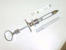
Dental syringe
A dental syringe is a syringe for the injection of a local anesthetic.[44] It consists of a breech-loading syringe fitted with a sealed cartridge containing anesthetic solution.
In the UK and Ireland, manually operated hand syringes to inject Lidocaine in to patient's gums.[45][46][44]
Other anesthetics used in dentistry
- Topical anesthetics benzocaine, eugenol, and forms of xylocaine are used topically to numb various areas before injections or other minor procedures.
- Nitrous oxide (N2O), also known as "laughing gas", easily crosses the alveoli of the lung and is dissolved into the passing blood, where it travels to the brain, leaving a dissociated and euphoric feeling in most cases. Nitrous oxide is used in combination with oxygen. Often (especially with children) a sweet-smelling fruity scent similar to an auto scent is used with the gas to inspire deep inhalation.
- General anesthesia drugs such as midazolam, ketamine, propofol and fentanyl are used to put a person in a twilight sleep or render them completely unconscious and unaware of pain. Dentists who have completed a training program in anesthesiology may also administer general IV and inhalation anesthetic agents.
- Nebotamine, a drug with similar effects to ketamine, is injected into the anterior lingual glands blocking action potentials from sending signals to the myelinated nerve. The potency of the anesthetic is directly related to its lipid solubility, since 90% of the nerve cell membrane is composed of lipid.
- Midazolam (Versed), a drug that represses memories of the procedure, is usually given two hours prior to the procedure in combination with Tylenol in general anesthesia so the person will go home with no memories of being in surgery.
- Sevoflurane gas in combination with nitrous oxide and oxygen is often used during general anesthesia followed by the use of isoflurane gas to maintain anesthesia during the procedure. In children sweet fruity scents are often used with the gases to inspire deep inhalation. Scents come in cherry, apple, bubblegum, watermelon, etc...
- Propofol, a drug with similar effects to Sodium Pentathol, is often used through intravenous infusion through an IV during general anesthesia after gasses are initiated.
- Morphine is often used to control pain during the dental surgery under general anesthesia. The morphine is usually administered through IV.
- Ketorolac is often administered through IV to suppress both pain and inflammation while under general anesthesia.
Other drugs used in combination with general anesthesia in dentistry
- Decadron a steroid is often administered through IV to suppress inflammation and swelling resulting during the surgery while under general anesthesia.
- Ondansetron brand named Zofran is often administered to prevent nausea during the surgery which may result from the blood draining into the stomach while under general anesthesia, or it is given after the procedure for postoperative nausea which may result from the anesthesia itself which was administered.
Local anesthesia and the pregnant patient
Provided a dentist performs proper aspiration to avoid intravenous injections, local anesthetics containing epinephrine (adrenaline) are safe to use during pregnancy. lignocaine and prilocaine are assigned a category B ranking by the FDA and are therefore safe for use during pregnancy. Lignocaine and prilocaine are sold as 2% and 4% formulations, respectively. It is therefore safer to use the lignocaine so as to administer a lower concentration of the drug to the pregnant patient.[47]
Mepivicaine, articaine, bupivicaine are given an FDA category C ranking and so should be avoided. Benzocaine, the ingredient of most topical anesthetic formulations, is also ranked as category C and should be avoided. Lignocaine should be used as topical anesthetic instead.[47]
Epinephrine in high doses is harmful to a pregnant woman in that it affects uterine blood flow. However its use in low dose with local anesthetic administration is warranted. The epinephrine causes vasoconstriction which in turn reduces systemic distribution of the anesthetic as well as prolongs its action in addition to decreasing bleeding at the operating site. Lidocaine 2% with 1:100,000 adrenaline is the local anesthetic of choice in the treatment of pregnant women.[47]
Allergy to local anaesthetic
Allergic reactions from local anaesthesia have been reported in some patients. However, this occurrence is rare even in patients who had a past history of adverse reactions to LA.
There are mainly 2 classes of local anaesthetic agents: Amide or Ester linkages, based on their chemical structure.[48]
- E.g. of amide LA: lidocaine, prilocaine, articaine, mepivacaine
- E.g. of ester LA: benzocaine, procaine
Genuine allergic reactions of an amide LA is very uncommon. An ester LA is more possible to result in an allergic reaction because the compound will be broken down to para-aminobenzoic acid (PABA) which is a trigger for allergic reactions.[49] In general dentistry, only topical applications of LA contains esters (benzocaine) when applied onto area before LA is administered.
If one is allergic to an ester LA, then the use of other types of ester LA should be avoided as the breakdown of all esters will produce PABA. However, patients allergic to ester LA will usually not be affected by amide LA because PABA is not produced upon breakdown of amide LA. Unlike ester LA, allergy to an amide LA will not eliminate the use of other types of amide LA.[49]
Some reactions are caused by administration of too much drug, usually because of the route of entry of drug (intravenously) or the quick uptake of drug into the system, or the aftereffect of the vasoconstrictor.[50] Unfavourable reactions to LA can be classified into 3 different groups: psychogenic, allergic, toxic.
Differential Diagnosis & management
- Psychogenic reactions
Unfavourable reactions to LA are commonly be due to a hyperemotional response to a perceived danger within someone’s mind, and it could be demonstrated in several ways. Examples are temporal loss of consciousness, sweating, flush, change in heart rate or blood pressure, panic attack, hyperventilation, of which may be mistaken as allergic reactions.
When treating such patients, treat them with care and take into consideration their anxiety. During treatment if the patients feel faint or experiences a drop in blood pressure, lay them flat and keep their legs elevated in an attempt to restore their blood pressure. Loosen any tight clothing and keep the patients sugary food/drink after they regain consciousness. Reassure the patient.
- Toxic reactions
This may occur when there are large amounts of anaesthetic within their vascular system, which may be due to them receiving repeated LA, intravenous entry of drug, or have underlying systemic conditions that does not metabolize or utilize the drug efficiently.[49] Signs and symptoms mainly involve the nervous system e.g. aggressive behaviour, drowsiness, speech alteration, disorientation etc.
Their symptoms should usually resolve in a few hours, up to 12 hours, as the body will gradually rid the bloodstream of the drug. Assure the patient that their symptoms will improve after a few hours and that such a reaction should not recur, and that there is no need to abstain from that drug hereafter.
Such reactions can be minimized via practicing safe injection methods using an aspirating syringe to prevent injecting in blood vessels, slow administration of drug, and avoid overprescribing LA, keeping in mind the patient’s weight, age and medical history.
Signs & symptoms of allergic reactions to LA
Genuine allergy to LA will manifest either as Type 1 or Type 4 hypersensitivity. Signs and symptoms will vary depending on the type of allergy. Type 1 reactions have a rapid onset of symptoms which include swelling, redness, rashes, itchiness, chest tightness, breathing problems. A Type 4 reaction has a delayed onset of symptoms and is usually localized to the site of injection.
Management
If a genuine allergic reaction to LA should occur, the patient should be treated as an emergency for anaphylaxis, according to the guidelines in the respective areas. For the UK, the section on medical emergencies in dental practice in the “Prescribing in Dental Practice” part in the BNF should be referred to. The patient should be sent immediately to the hospital if their condition worsens.[49]
The individual should undergo further tests to certify their allergy to the LA or for other possible causes of the adverse reaction.
Gate Control Theory in Painless Anaesthesia
The gate control theory explains that pain can be reduced if the touch nerve fibres are stimulated due to non-harmful stimuli.
Advancement in techniques used to deliver local anaesthesia are very important. There are types of local anaesthesia that apply vibrations to the skin while the injection is being placed into the skin. This uses the gate control theory to minimise pain to the patient. The high frequency vibrations coming from the device which is attached to the syringe inhibit the pain sensations coming from the needle. They may interfere with the signals of pain by closing the gate in the brain. The nerve fibres that are stimulated are the Ab fibres using pressure or vibration. Other receptors called meissner’s corpuscles in the deeper tissues and bone also contribute. This closes a ‘neural gate’ This decreases the patient’s feeling of pain.
Methods used by dentist to reduce pain during anaesthesia by using the gate control theory are: Warming of the local anaesthetic cartridge, Stretching the oral mucosa, Gentle rubbing of the extra-oral skin.[51]
See also
References
- 1 2 3 4 5 6 7 8 9 10 11 12 13 14 15 16 17 18 19 G., Meechan, J. (2010). Practical dental local anaesthesia. Wilson, Nairn H. F. (2nd ed.). London: Quintessence Pub. Co. ISBN 1850972044. OCLC 642291828.
- 1 2 3 4 5 "Oral Nerve Block: Overview, Indications, Contraindications". 2018-02-01.
- ↑ Khan SR, Qazi SR (November 2017). "Extraction of maxillary teeth by dental students without palatal infiltration of local anaesthesia: a randomised controlled trial". European Journal of Dental Education. 21 (4): e39–e42. doi:10.1111/eje.12215. PMID 27324934.
- 1 2 Khalil H (2014). "A basic review on the inferior alveolar nerve block techniques". Anesthesia, Essays and Researches. 8 (1): 3–8. doi:10.4103/0259-1162.128891. PMC 4173572. PMID 25886095.
- ↑ Palti DG, Almeida CM, Rodrigues Ade C, Andreo JC, Lima JE (2011). "Anesthetic technique for inferior alveolar nerve block: a new approach". Journal of Applied Oral Science. 19 (1): 11–5. doi:10.1590/S1678-77572011000100004. PMC 4245857. PMID 21437463.
- ↑ Buch HA (May 2011). "Clinical anatomy of inferior alveolar nerve block anesthesia". Clinical Anatomy. 24 (4): 515–7. doi:10.1002/ca.21136. PMID 21374723.
- ↑ B.,, Bassett, Kathy. Local anesthesia for dental professionals. DiMarco, Arthur C.,, Naughton, Doreen K., (Second ed.). Pearson. ISBN 9780133077711. OCLC 875056114.
- ↑ Nicholson ML (May 1985). "A study of the position of the mandibular foramen in the adult human mandible". The Anatomical Record. 212 (1): 110–2. doi:10.1002/ar.1092120116. PMID 4073538.
- ↑ Tzermpos FH, Cocos A, Kleftogiannis M, Zarakas M, Iatrou I (2012). "Transient delayed facial nerve palsy after inferior alveolar nerve block anesthesia". Anesthesia Progress. 59 (1): 22–7. doi:10.2344/11-03.1. PMC 3309298. PMID 22428971.
- ↑ Ethunandan M, Tran AL, Anand R, Bowden J, Seal MT, Brennan PA (April 2007). "Needle breakage following inferior alveolar nerve block: implications and management". British Dental Journal. 202 (7): 395–7. doi:10.1038/bdj.2007.272. PMID 17435721.
- ↑ Yoon RK, Chussid S (July 2012). "Ocular complications following an inferior alveolar nerve block on a child patient: a review of the literature and report of a case". Pediatric Dentistry. 34 (4): 343–6. PMID 23014093.
- ↑ Drum M, Reader A, Beck M (July 2011). "Long buccal nerve block injection pain in patients with irreversible pulpitis". Oral Surgery, Oral Medicine, Oral Pathology, Oral Radiology, and Endodontics. 112 (1): e51–4. doi:10.1016/j.tripleo.2011.01.028. PMID 21458333.
- ↑ Foster W, Drum M, Reader A, Beck M (2007). "Anesthetic efficacy of buccal and lingual infiltrations of lidocaine following an inferior alveolar nerve block in mandibular posterior teeth". Anesthesia Progress. 54 (4): 163–9. doi:10.2344/0003-3006(2007)54[163:AEOBAL]2.0.CO;2. PMC 2213247. PMID 18085837.
- ↑ Yeşilyurt H, Aydinlioglu A, Kavakli A, Ekinci N, Eroglu C, Hacialiogullari M, Diyarbakirli S (2008). "Local differences in the position of the mental foramen". Folia Morphologica. 67 (1): 32–5. PMID 18335411.
- ↑ Santini A, Land M (1990). "A comparison of the position of the mental foramen in Chinese and British mandibles". Acta Anatomica. 137 (3): 208–12. PMID 2349864.
- ↑ Shankland WE (1994). "The position of the mental foramen in Asian Indians". The Journal of Oral Implantology. 20 (2): 118–23. PMID 7869414.
- ↑ Masselink, B.H. (1910). "The advent of painless dentistry". Dent Cosmo. 52 (8): 868–872.
- ↑ Gallatin J, Reader A, Nusstein J, Beck M, Weaver J (November 2003). "A comparison of two intraosseous anesthetic techniques in mandibular posterior teeth". Journal of the American Dental Association. 134 (11): 1476–84. PMID 14664266.
- 1 2 1944-, Malamed, Stanley F., (2013). Handbook of local anesthesia (6th ed.). St. Louis, Missouri: Elsevier. ISBN 9780323074131. OCLC 769141511.
- 1 2 Claffey E, Reader A, Nusstein J, Beck M, Weaver J (August 2004). "Anesthetic efficacy of articaine for inferior alveolar nerve blocks in patients with irreversible pulpitis". Journal of Endodontics. 30 (8): 568–71. PMID 15273637.
- ↑ Malamed, SF (1982). "The PDL injection: An alternative to inferior alveolar nerve block". PMID 6949113.
- ↑ "Supplemental injections" (PDF). columbia.edu.
- ↑ Pradhan, Deepak, Lakshmi, Raunak, Kulkarni, Shetty (January 2011). "Evaluation of Efficacy of Intraligamentary Injection Technique for Extraction of Mandibular Teeth-A Prospective Study".
- ↑ "supplemental injections" (PDF).
- ↑ "Supplementary techniques". pocketdentistry.com.
- 1 2 Illustrated Anatomy of the Head and Neck, Fehrenbach and Herring, Elsevier, 2012, page 216
- 1 2 3 4 Akinosi, J.O. (1977). "A new approach to the mandibular nerve block". British Journal of Oral Surgery. 15 (1): 83–87.
- 1 2 3 4 Lenka, Sthitaprajna & Jain Kumar, Nikil & Mohanty, Rajat & Singh, Dhirendra & Gulati, Minkle (2013). "A Clinical Comparison of Three Techniques of Mandibular Local Anaesthesia". Journal of Research and Advancement in Dentistry. 2: 61–67.
- 1 2 3 4 5 6 7 8 Katch EM (May–June 1986). "Application of transcutaneous electrical nerve stimulation in dentistry". Anesthesia Progress. American Dental Society of Anesthesiology. 33 (3): 156–60. eISSN 1878-7177. PMC 2175474. PMID 3488697.
- 1 2 3 4 5 6 7 8 Vikrant Kasat, Aditi Gupta, Ruchi Ladda, Mitesh Kathariya, Harish Saluja, and Anjum-Ara Farooqui (Dec 2014). "Transcutaneous electric nerve stimulation (TENS) in dentistry- A review". J Clin Exp Dent. PMC 4312687.
- 1 2 3 4 5 6 7 8 "Transcutaneous Electrical Nerve Stimulation". 9 December 2015. Retrieved 10 February 2018.
- 1 2 3 4 "TENS (transcutaneous electrical nerve stimulation)". 6 October 2015. Retrieved 10 February 2018.
- 1 2 "Advances in dental local anesthesia techniques and devices: An update". Advances in dental local anesthesia techniques and devices: An update. 2013 Jan-Jun. PMC 3800379. Check date values in:
|date=(help) - ↑ "Dentium | Product | New Product". www.dentium.com. Retrieved 2018-08-04.
- ↑ "Articaine / Epinephrine Dosage". Retrieved 10 February 2018.
- ↑ "Local Anaesthetic". Retrieved 7 February 2018.
- ↑ "Maximum Recommended Local Anaesthetic Doses for Adults". Retrieved 10 February 2018.
- ↑ "Scandonest 3% Plain". Retrieved 10 February 2018.
- ↑ https://books.google.co.uk/books?id=xRgnDwAAQBAJ&pg=PA52&dq=dental+anaesthesia+contraindications&hl=en&sa=X&ved=0ahUKEwjkmOnbmqHZAhUQ2qQKHX2DAUEQ6AEIJzAA#v=onepage&q=dental%20anaesthesia%20contraindications&f=false
- ↑ Tong, Huei Jinn; Alzahrani, Fatma Salem; Sim, Yu Fan; Tahmassebi, Jinous F.; Duggal, Monty (2018-04-10). "Anaesthetic efficacy of articaine versus lidocaine in children's dentistry: a systematic review and meta-analysis". International Journal of Paediatric Dentistry. doi:10.1111/ipd.12363. ISSN 1365-263X. PMID 29635712.
- ↑ https://books.google.co.uk/books?id=dmxjDQAAQBAJ&pg=PA151&dq=dental+anaesthesia+contraindications&hl=en&sa=X&ved=0ahUKEwjkmOnbmqHZAhUQ2qQKHX2DAUEQ6AEILDAB#v=onepage&q=dental%20anaesthesia%20contraindications&f=false
- 1 2 Local Anesthesia for the Dental Hygienist, Logothetis, Elsevier, 2012
- ↑ Local Anesthesia for the Dental Hygienist, Logothetis, Elsevier, 2012
- 1 2 "Lidocaine Hydrochloride (Local) Monograph for Professionals - Drugs.com".
- ↑ Zakrzewska JM, Boon EC (August 2003). "Use of safety dental syringes in British and Irish dental schools". British Dental Journal. 195 (4): 207–9, discussion 198. doi:10.1038/sj.bdj.4810445. PMID 12970703 – via www.nature.com.
- ↑ Zakrzewska JM, Greenwood I, Jackson J (27 January 2001). "Cross-infection control: Introducing safety syringes into a UK dental school – a controlled study". British Dental Journal. 190 (2): 88–92. doi:10.1038/sj.bdj.4800891 – via www.nature.com.
- 1 2 3 Ouanounou A, Haas DA (April 2016). "Drug therapy during pregnancy: implications for dental practice". British Dental Journal. 220 (8): 413–7. doi:10.1038/sj.bdj.2016.299. PMID 27103292.
- ↑ Haas, D. A (2002). "An update on local anesthetics in dentistry". Journal (Canadian Dental Association).
- 1 2 3 4 Henderson, S. (2011). "Allergy to local anaesthetic agents used in dentistry--what are the signs, symptoms, alternative diagnoses and management options?". Dental Update. 38 (6): 410–2. PMID 21905354.
- ↑ Tomoyasu, Y., Mukae, K., Suda, M., Hayashi, T., Ishii, M., Sakaguchi, M., … Miyawaki, T. (2011). "Allergic reactions to local anesthetics in dental patients: analysis of intracutaneous and challenge tests". The Open Dentistry Journal. 5: 146–9.
- ↑ Technique tips--distraction anaesthesia: applying the gate control theory in delivering painless anaesthesia. Malik A.
External links
- Endo T, Gabka J, Taubenheim L (January 2008). "Intraligamentary anesthesia: benefits and limitations". Quintessence International. 39 (1): e15–25. PMID 18551207.
| Types | |
|---|---|
| Techniques | |
| Scientific principles | |
| Measurements | |
| Instruments | |
| Complications | |
| Fields of study | |
| Professions | |
| History |
|
| Organizations |
|
| |