Cyanine
Cyanine is the non-systematic name of a synthetic dye family belonging to polymethine group. The word cyanin is from the English word "cyan", which conventionally means a shade of blue-green (close to "aqua") and is derived from the Greek κυάνεος/κυανοῦς kyaneos/kyanous which means a somewhat different color: "dark blue".
Cyanines were and are still used in industry, and more recently in biotechnology (labeling, analysis). Cyanines have many uses as fluorescent dyes, particularly in biomedical imaging. Depending on the structure, they cover the electromagnetic spectrum from near IR to UV. There are a large number reported in the literature.
Structure
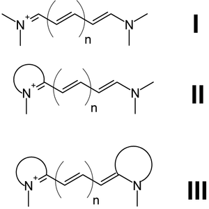
I = Streptocyanines,
II = Hemicyanines,
III = Closed cyanines
There are three types of cyanines:[1]
- Streptocyanines or open chain cyanines:
- R2N+=CH[CH=CH]n-NR2 (I)
- Hemicyanines:
- Aryl=N+=CH[CH=CH]n-NR2 (II)
- Closed chain cyanines:
- Aryl=N+=CH[CH=CH]n-N=Aryl (III)
where two quaternary nitrogens are joined by a polymethine chain.[2] Both nitrogens may each be independently part of a heteroaromatic moiety, such as pyrrole, imidazole, thiazole, pyridine, quinoline, indole, benzothiazole, etc.
History and Use in Industry
Cyanines were first synthesized over a century ago. They were originally used, and still are, to increase the sensitivity range of photographic emulsions, i.e., to increase the range of wavelengths which will form an image on the film, making the film panchromatic. Cyanines are also used in CD-R and DVD-R media. The ones used are mostly green or light blue in color, and are chemically unstable. This makes unstabilized cyanine discs unsuitable for archival CD and DVD use, as they can fade and become unreadable in a few years, however, recent cyanine discs contain stabilizers that slow down the deterioration significantly. These discs are often rated with an archival life of 75 years or more. The other dyes used in CD-Rs are phthalocyanine and azo.
Use in Biotechnology
Cyanines dyes are usually synthesized from 2, 3, 5 or 7-methine structures with reactive groups on either one or both of the nitrogen ends so that they can be chemically linked to either nucleic acids or protein molecules. Labeling is done for visualization and quantification purposes. Biological applications include comparative genomic hybridization and gene chips, which are used in transcriptomics, and various studies in proteomics such as RNA localization,[3] molecular interaction studies by fluorescence energy transfer (FRET) and fluorescent immunoassays.
Alan S. Waggoner et al of Carnegie-Mellon University filed a key patent application in this field in 1986, which later resulted in the grant of a number of patents relating to the use of Cyanine dyes including US6225050 (B1) in 2001 and US6956032 (B1) in 2005. This intellectual property was licensed to GE Healthcare.
In addition GE Healthcare registered "Cy" and "CyDye" as trademarks.
Cyanines dyes are available with different modifications such as methyl, ethyl or butyl substituents, carboxyl, acetylmethoxy, and sulfo groups which alter their hydrophilicity.[4]
| Probe | Ex (nm) | Em (nm) | MW | Quantum yield |
|---|---|---|---|---|
| Cy2 | 489 | 506 | 714 | QY 0.12 |
| Cy3 | (512);550 | 570;(615) | 767 | QY 0.15 [5] |
| Cy3B | 558 | 572;(620) | 658 | QY 0.67 |
| Cy3.5 | 581 | 594;(640) | 1102 | QY 0.15 |
| Cy5 | (625);650 | 670 | 792 | QY 0.27[5] |
| Cy5.5 | 675 | 694 | 1128 | QY 0.28[6] |
| Cy7 | 743 | 767 | 818 | QY 0.28 |
Ex (nm): Excitation wavelength in nanometers
Em (nm): Emission wavelength in nanometers
MW: Molecular weight
QY: Quantum yield
Common Cyanine dyes and their uses
Cyanines can advantageously replace conventional dyes such as fluorescein and rhodamines because they yielding brighter and more stable fluorescence.
- Cy3 and Cy5 are the most popular, used typically combined for 2 colors detection.
Cy3 fluoresces greenish yellow (~550 nm excitation, ~570 nm emission), while Cy5 is fluorescent in the red region (~650 excitation, 670 nm emission).[7] Cy3 can be detected by various fluorometers, imagers, and microscopes with standard filters for Tetramethylrhodamine (TRITC). Due to its high molar extinction coefficient, this dye is also easily detected by naked eye on electrophoresis gels, and in solution. Cy5 became a popular replacement for far red fluorescent dyes because of its high extinction coefficient (as small as 1 nanomol can be detected in gel electrophoresis by naked eye) and its fluorophore emission maximum in the red region, where many CCD detectors have maximum sensitivity and biological objects give low background interference.
The scanners actually use different laser emission wavelengths (typically 532 nm and 635 nm) and filter wavelengths (550-600 nm and 655-695 nm) to avoid background contamination. They are thus able to easily distinguish colors from Cy3 and from Cy5, and also able to quantify the amount of Cy3 and Cy5 labeling in one sample (multiparametric detection).
- Other cyanine dyes are useful:
Cy3.5 can replace SulfoRhodamine 101.
Cy5.5 is a near-infrared (IR) fluorescence-emitting dye (Excitation/emission maximum 678/694 nm).
Cy7 is a near-IR fluor that is invisible to the naked eye (Excitation/emission maximum 750/776 nm). It is used in in vivo imaging applications, as well as the Cy7.5 dye.
Sulfo–Cyanine dyes bear one or two sulfo groups, rendering the Cy dye water-soluble, but tri- and quadri-sulfonated forms are available for even higher water solubility.[4] PEGylation is another modification that confers hydrophilicity, not only to the dye but also to the labeled conjugate.
Nomenclature and Structure
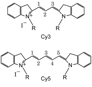
Standard chemical names exactly specify the chemical structure of molecules. The Cy3 and Cy5 nomenclature was first proposed by Ernst, et al.[2] in 1989, and is non-standard since it gives no hint of their chemical structures. In the original paper the number designated the count of the methines (as shown), and the side chains were unspecified. Due to this ambiguity various structures are designated Cy3 and Cy5 in the literature.
The R groups do not have to be identical. In the dyes as used they are short aliphatic chains one or both of which ends in a highly reactive moiety such as N-hydroxysuccinimide or maleimide.
Cyanine Dye Alternatives
Many analogs of standard Cy 2 / 3 / 3.5 / 5 / 5.5 / 7 / 7.5 dyes were developed, using diverse modification: Alexa Fluor dyes, Dylight, FluoProbes dyes, Sulfo Cy dyes,[8] Seta dyes,[9] IRIS dyes from Cyanine Technologies [10] and others can be used interchangeably with Cy dyes in most biochemical applications, with claimed improvements in solubility, fluorescence, or photostability.[11][12]
Whilst patent based IP protection for the standard Cy series of dyes has now lapsed, trademark based IP continues to inhibit the promotion of dyes with the original Cy naming. As a result dyes that are chemically identical to Cy dyes, but called different names, are now sold.
Cy5 ozone susceptibility
In 2003, researchers at Inpharmatics and Agilent reported in Analytical Chemistry that microarrays which used Cy5 were susceptible to intermittent data quality decrease caused by environmental ozone. Exposures to ozone levels above 5-10 ppb for 10–30 seconds were reported to decrease the reproducibility of Cy5 microarrays. Much higher levels of ozone (>100 ppb) were required to observe an effect in Cy3.[13] There are devices that claim to remove ambient ozone levels but they have not been 3rd party tested.
Applications

Cyanine dyes are used to label proteins, antibodies, peptides, nucleic acid probes, and any kind of other biomolecules to be used in a variety of fluorescence detection techniques: Flow cytometry, Microscopy (mainly Visible range, but also UV, IR), Microplate assays, Microarrays, as well as "light-up Probes". [14]
Nucleic Acid Labeling
In microarray experiments DNA or RNA is labeled with either Cy3 or Cy5 that has been synthesized to carry an N-hydroxysuccinimidyl ester (NHS-ester) reactive group. Since NHS-esters react readily only with aliphatic amine groups, which nucleic acids lack, nucleotides have to be modified with aminoallyl groups. This is done through incorporating aminoallyl-modified nucleotides during synthesis reactions. A good ratio is a label every 60 bases such that the labels are not too close to each other, which would result in quenching effects.
Protein labeling
For protein labeling, Cy3 and Cy5 dyes sometimes bear a succinimidyl group to react with amines, or a maleimide group to react with a sulfhydryl group of cysteine residues.
Cy5 is sensitive to the electronic environment it resides in. Changes in the conformation of the protein it is attached to will produce either enhancement or quenching of the emission. The rate of this change can be measured to determine enzyme kinetic parameters. The dyes can be used for similar purposes in FRET experiments.
Cy3 and Cy5 are used in proteomics experiments so that samples from two sources can be mixed and run together through the separation process.[15][16] This eliminates variations due to differing experimental conditions that are inevitable if the samples were run separately. These variations make it extremely difficult, if not impossible, to use computers to automate the acquisition of the data after the separation is complete. Using these dyes makes the automation trivial.
References
- ↑ Kim, Eunha; Park, Seung Bum (2010). "Discovery of New Synthetic Dyes: Targeted Synthesis or Combinatorial Approach?". In Demchenko, Alexander P. Advanced Fluorescence Reporters in Chemistry and Biology I: Fundamentals and Molecular Design Volume 8 of Springer Series on Fluorescence. Berlin: Springer. p. 172. ISBN 9783642047022.
- 1 2 Ernst LA, Gupta RK, Mujumdar RB, Waggoner AS (Jan 1989). "Cyanine dye labeling reagents for sulfhydryl groups". Cytometry. 10 (1): 3–10. doi:10.1002/cyto.990100103. PMID 2917472.
- ↑ Blower MD, Feric E, Weis K, Heald R (Dec 2007). "Genome-wide analysis demonstrates conserved localization of messenger RNAs to mitotic microtubules". The Journal of Cell Biology. 179 (7): 1365–73. doi:10.1083/jcb.200705163. PMC 2373496. PMID 18166649.
- 1 2 CYanine dyes
- 1 2 Mujumdar B, Ernst A, Mujumdar SR, Lewis CJ, Waggoner AS (Mar 1993). "Cyanine dye labeling reagents: Sulfoindocyanine succinimidyl esters". Bioconjugate Chemistry. 4 (2): 105–111. doi:10.1021/bc00020a001.
- ↑ Umezawa K, Matsui A, Nakamura Y, Citterio D, Suzuki K (2009). "Bright, color-tunable fluorescent dyes in the Vis/NIR region: establishment of new "tailor-made" multicolor fluorophores based on borondipyrromethene". Chemistry. 15 (5): 1096–106. doi:10.1002/chem.200801906. PMID 19117043.
- ↑ Jackson ImmunoResearch. "Cyanine Dyes (Cy2, Cy3, and Cy5)". Retrieved 2008-10-31.
- ↑ Cyandye, LLC
- ↑ SETA BioMedicals
- ↑ "Archived copy". Archived from the original on 2015-01-26. Retrieved 2015-01-26.
- ↑ FluoProbes488 comparison to FITC, Cyanine2
- ↑ FluoProbes547H comparison in Confocal Microscopy
- ↑ Fare TL, Coffey EM, Dai H, He YD, Kessler DA, Kilian KA, Koch JE, LeProust E, Marton MJ, Meyer MR, Stoughton RB, Tokiwa GY, Wang Y (Sep 2003). "Effects of atmospheric ozone on microarray data quality". Analytical Chemistry. 75 (17): 4672–5. doi:10.1021/ac034241b. PMID 14632079.
- ↑ Armitage, Bruce A. (27 January 2005). "Cyanine Dye–DNA Interactions: Intercalation, Groove Binding, and Aggregation". Topics in Current Chemistry. Springer Berlin Heidelberg: 55–76. doi:10.1007/b100442.
- ↑ Unlü M, Morgan ME, Minden JS (Oct 1997). "Difference gel electrophoresis: a single gel method for detecting changes in protein extracts". Electrophoresis. 18 (11): 2071–7. doi:10.1002/elps.1150181133. PMID 9420172.
- ↑ Osterman IA, Ustinov AV, Evdokimov DV, Korshun VA, Sergiev PV, Serebryakova MV, Demina IA, Galyamina MA, Govorun VM, Dontsova OA (Jan 2013). "A nascent proteome study combining click chemistry with 2DE" (PDF). Proteomics. 13 (1): 17–21. doi:10.1002/pmic.201200393. PMID 23161590.
External links
| Look up cyanine in Wiktionary, the free dictionary. |