Common peroneal nerve
| Common peroneal nerve | |
|---|---|
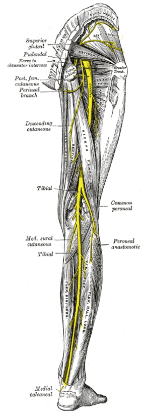 Nerves of the right lower extremity Posterior view. (Common peroneal labeled at center right.) | |
| Details | |
| From | sacral plexus via sciatic nerve (L4-S2) |
| To | Deep fibular nerve and Superficial fibular nerve |
| Innervates | Anterior compartment of leg, lateral compartment of leg, extensor digitorum brevis |
| Identifiers | |
| Latin |
Nervus fibularis communis, Nervus peronaeus communis |
| MeSH | D010543 |
| TA | A14.2.07.047 |
| FMA | 19039 |
| Anatomical terms of neuroanatomy | |
The common peroneal nerve (common fibular nerve; external popliteal nerve; lateral popliteal nerve) is a nerve in the lower leg that provides sensation over the posterolateral part of the leg and the knee joint. Its is divided into terminal branches of superficial peroneal nerve and deep peroneal nerve, where the latter of the two supplies the muscles of the anterior and lateral compartments of the leg. When the common peroneal nerve is damaged or compressed, foot drop can be the end result.
Structure
The common peroneal nerve is the smaller terminal branch of the sciatic nerve. The common peroneal nerve has root values of L4, L5, S1, and S2. It arises from the superior angle of the popliteal fossa and extends to the lateral angle of the popliteal fossa, along the medial border of the biceps femoris. It then winds around the neck of the fibula to pierce the peroneus longus and divides into terminal branches of superficial peroneal nerve and deep peroneal nerve. Before its division, the common peroneal nerve gives off several branches in the popliteal fossa.[1]
Cutaneous branches
- Lateral sural cutaneous nerve (lateral cutaneous nerve of calf) - supplies the skin of the upper two-thirds of the lateral side of leg.[1]
- peroneal communicating nerve - it runs on the posterolateral aspect of the calf and joins the sural nerve.[1]
Articular branches
- Superior lateral genicular nerve - accompanies artery of the same name and lies above the lateral femoral condyle.[1]
- Inferior lateral genicular nerve - accompanies artery of the same name and lies just above the head of the fibula.[1]
- Recurrent genicular nerve - It arises from the point of division of the common peroneal nerve; then ascends anterior to the knee joint together with the anterior recurrent tibial artery to supply the knee joint and the tibialis anterior muscle.[1]
Motor branches
There are no motor branches arising directly from common peroneal nerve.[1]
Function
The common peroneal nerve innervates the short head of the biceps femoris muscle via a motor branch that exits close to the gluteal cleft.[2] The remainder of the peroneal-innervated muscles are innervated by its branches, the deep peroneal nerve and superficial peroneal nerve.
It provides sensory innervation to the skin over the upper third of the lateral aspect of the leg via the lateral cutaneous nerve of the calf.[2] It gives the peroneal communicating nerve which joins the sural nerve in the midcalf.
Clinical significance
Chronic peroneal neuropathy can result from, among other conditions, bed rest of long duration, hyperflexion of the knee, peripheral neuropathy, pressure in obstetric stirrups, and conditioning in ballet dancers. The most common cause is habitual leg crossing that compresses the common peroneal nerve as it crosses around the head of the fibula.[3] Transient trauma to the nerve can result from peroneal strike.
Damage to this nerve typically results in foot drop, where dorsiflexion of the foot is compromised and the foot drags (the toe points) during walking; and in sensory loss to the dorsal surface of the foot and portions of the anterior, lower-lateral leg. A common yoga kneeling exercise, the Vajrasana, has been linked to a variant called yoga foot drop.[4][5]
Surgical procedures involving the nerve involve:
- Peroneal nerve decompression
- Deep peroneal nerve decompression
- In the surgical treatment of deep peroneal nerve entrapment in the foot, a ligament from the extensor digitorum brevis muscle that crosses over the deep peroneal nerve, putting pressure on it and causing pain, is released.[9]
Additional images
 Front and posterior views of cutaneous nerves of the right lower extremity
 Common fibular nerve
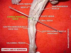 Common peroneal nerve
 Common peroneal nerve
See also
References
This article incorporates text in the public domain from page 964 of the 20th edition of Gray's Anatomy (1918)
- 1 2 3 4 5 6 7 Krishna, Garg (2010). "Popliteal fossa (Chapter 6)". BD Chaurasia's Human Anatomy (Regional and Applied Dissection and Clinical) Volume 2 - Lower limb, abdomen, and pelvis (Fifth ed.). India: CBS Publishers and Distributors Pvt Ltd. p. 87,88. ISBN 978-81-239-1864-8.
- 1 2 Katirji, Bashar (2007). Electromyography in Clinical Practice: A Case Study Approach, 2nd ed. Mosby Elsevier. p. 146. ISBN 9780323028998.
- ↑ Bradley, Walter G.; et al. (2004). Neurology in Clinical Practice (4th ed.). Philadelphia: Butterworth-Heinemann. pp. 453–454. ISBN 0-7506-7469-5.
- ↑ Joseph Chusid (August 9, 1971). "Yoga Foot Drop". JAMA: The Journal of the American Medical Association. 271 (6): 827–828. doi:10.1001/jama.1971.03190060065025. Retrieved November 19, 2012.
- ↑ William J. Broad (January 5, 2012). "How Yoga Can Wreck Your Body". The New York Times Magazine. Retrieved August 29, 2012.
- ↑ "Peroneal Nerve Injury (Foot Drop)", Neurology and Neurosurgery, Johns Hopkins School of Medicine, 2013-12-18, retrieved 2013-12-18
- ↑ "Peroneal Nerve Entrapment at the Fibular Head", Department of Neurosurgery, NYU Langone Medical Center, 2013-12-18, retrieved 2013-12-18
- ↑ About us, Dellon Institutes for Peroneal Nerve Surgery, 2013-12-18, retrieved 2013-12-18
- 1 2 Dellon Institutes Peroneal Nerve Compression Surgical Treatment (pdf), Dellon Institutes for Peroneal Nerve Surgery, 2006-02-20, retrieved 2013-12-18
External links
- Anatomy photo:14:st-0501 at the SUNY Downstate Medical Center
- Peroneal_nerve at the Duke University Health System's Orthopedics program
- latleg at The Anatomy Lesson by Wesley Norman (Georgetown University)
- arteries-nerves%20LE/nerves4 at the Dartmouth Medical School's Department of Anatomy
- Overview at okstate.edu