Écorché

.jpg)
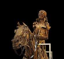
An écorché (French pronunciation: [ekɔʁʃe]) is a figure drawn, painted, or sculpted showing the muscles of the body without skin, normally as a figure study for another work or as an exercise for a student artist. The Renaissance-era architect, theorist and all-around Renaissance man, Leon Battista Alberti, recommended that when painters intend to depict a nude, they should first arrange the muscles and bones, then depict the overlying skin.[1]
Some of the first well known studies of this kind were performed by Leonardo da Vinci, who dissected cadavers and created detailed drawings of them. However, there are some accounts of this same practice taking place as far back as ancient Greece, though the specifics are not known.
Etymology
The term écorché, meaning literally "flayed", came into usage via the French Academies (such as the École des Beaux Arts) in the 19th century.[1]
History
Although there are some accounts of practices similar to écorché as far back as ancient Greece, the degree of similarity is unclear. The term as used today can be applied with the greatest confidence to the Renaissance period onwards.
Renaissance
During the Renaissance in Italy, around the 1450 to 1600, the rebirth of classical Greek and Roman characteristics in art led to the studies of the human anatomy. The practice of dissecting the human body was banned for many centuries due to the belief that body and soul were inseparable. It wasn’t until the election of Pope Boniface VIII that the practice of dissection was once again allowed for observation.[2][3] Many painters and artists documented and even performed the dissections themselves by taking careful observations of the human body. Among them were Leonardo da Vinci and Andreas Vesalius, two of the most influential artists in anatomical illustrations.[4] Leonardo da Vinci, in particular, was very detailed in his studies that he was known as the “artist-anatomist” for the creation of a new science and the creative depiction to anatomy. Leonardo’s anatomical studies contributed to artistic exploration of the movement of the muscles, joints and bones. His goal was to analyze and understand the instruments behind the postures and gestures in the human body.[5]
17th – 19th centuries
The study of anatomical figures became popular among the medical academies across Europe around the 17th and 18th century, especially when there was a lack of bodies available for dissections.[4] Medical students relied on these figures because they provided a good representation of what the anatomical model looks like. The écorché (flayed) figures were made to look like the skin was removed from the body, exposing the muscles and vessels of the model. Some figures were created to strip away the layers of muscles and reveal the skeleton of the model. Many of the life-size scale écorché figures were reproduced in a smaller scale out of bronze that could be easily distributed.[6]
Écorché figures were commonly made out of many different materials: bronze, ivory, plaster, wax, or wood. By the late eighteenth and early nineteenth centuries, wax was the most popular use of material in creating écorché statues. The production of colored wax anatomies allowed for a variety of hues and tone that makes the models appear realistic.[7]
21st century
The écorché form of study still continues at traditional schools throughout the world including the New York Academy of Art, the Art Students League of New York, the Grand Central Academy of Art in New York City, the Pennsylvania Academy of the Fine Arts in Philadelphia, and the Academy of Art University in San Francisco.[8]
References
- 1 2 Écorché defined at ArtLex.com
- ↑ Lemire, M (1 December 1992). "Representation of the human body: the colored wax anatomic models of the 18th and 19th centuries in the revival of medical instruction". Surgical and Radiologic Anatomy. 14 (4): 283–291. doi:10.1007/BF01794751. Retrieved 3 April 2013.
- ↑ Ginn, Sheryl R.; Lorusso, Lorenzo (16 July 2008). "Brain, Mind, and Body: Interactions with Art in Renaissance Italy". Journal of the History of the Neurosciences. 17 (3): 295–313. doi:10.1080/09647040701575900. PMID 18629698.
- 1 2 Wallace, Martin Kemp, Marina (2001). Spectacular bodies : the art and science of the human body from Leonardo to now. London: Hayward Gallery. pp. 22–90. ISBN 0520227921.
- ↑ Keele, Kenneth D. (October 1964). "Leonardo Da Vinci's Influence on Renaissance Anatomy". Medical History. 8 (4): 360–370. doi:10.1017/s0025727300029835. PMC 1033412. PMID 14230140.
- ↑ Owen, Harry (1 April 2012). "Early Use of Simulation in Medical Education". Simulation in Healthcare: the Journal of the Society for Simulation in Healthcare. 7 (2): 102–116. doi:10.1097/SIH.0b013e3182415a91.
- ↑ DARLINGTON, ANNE (1 December 1986). "The Teaching of Anatomy and the Royal Academy of Arts 1768-1782". Journal of Art & Design Education. 5 (3): 263–271. doi:10.1111/j.1476-8070.1986.tb00207.x.
- ↑ "Academy of Art University On Campus Labs" (PDF). academyart.edu. Academy of Art University. Retrieved 23 November 2016.
External links
![]()