Retrovirus
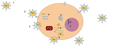
A retrovirus is a type of RNA virus[lower-alpha 1] that inserts a copy of its genome into the DNA of a host cell that it invades, thus changing the genome of that cell.[3] Once inside the host cell's cytoplasm, the virus uses its own reverse transcriptase enzyme to produce DNA from its RNA genome, the reverse of the usual pattern, thus retro (backwards). The new DNA is then incorporated into the host cell genome by an integrase enzyme, at which point the retroviral DNA is referred to as a provirus. The host cell then treats the viral DNA as part of its own genome, transcribing and translating the viral genes along with the cell's own genes, producing the proteins required to assemble new copies of the virus.
| Retroviruses | |
|---|---|
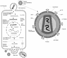 | |
| HIV retrovirus schematic of cell infection, virus production and virus structure | |
| Virus classification | |
| (unranked): | Virus |
| Phylum: | incertae sedis |
| Class: | incertae sedis |
| Order: | Ortervirales |
| Family: | Retroviridae |
| Subfamilies and genera[1] | |
| |
Human retroviruses include HIV-1 and HIV-2, the cause of the disease AIDS. Retroviruses are also a valuable research tools in molecular biology, and they have been used successfully in gene delivery systems.[4]
Structure
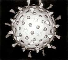
Virions of retroviruses consist of enveloped particles about 100 nm in diameter. The virions also contain two identical single-stranded RNA molecules 7–10 kilobases in length. Although virions of different retroviruses do not have the same morphology or biology, all the virion components are very similar.[5]
The main virion components are:
- Envelope: composed of lipids (obtained from the host plasma membrane during the budding process) as well as glycoprotein encoded by the env gene. The retroviral envelope serves three distinct functions: protection from the extracellular environment via the lipid bilayer, enabling the retrovirus to enter/exit host cells through endosomal membrane trafficking, and the ability to directly enter cells by fusing with their membranes.
- RNA: consists of a dimer RNA. It has a cap at the 5' end and a poly(A) tail at the 3' end. The RNA genome also has terminal noncoding regions, which are important in replication, and internal regions that encode virion proteins for gene expression. The 5' end includes four regions, which are R, U5, PBS, and L. The R region is a short repeated sequence at each end of the genome used during the reverse transcription to ensure correct end-to-end transfer in the growing chain. U5, on the other hand, is a short unique sequence between R and PBS. PBS (primer binding site) consists of 18 bases complementary to 3' end of tRNA primer. L region is an untranslated leader region that gives the signal for packaging of the genome RNA. The 3' end includes 3 regions, which are PPT (polypurine tract), U3, and R. The PPT is a primer for plus-strand DNA synthesis during reverse transcription. U3 is a sequence between PPT and R, which serves as a signal that the provirus can use in transcription. R is the terminal repeated sequence at 3' end.
- Proteins: consisting of gag proteins, protease (PR), pol proteins, and env proteins.
- Group-specific antigen (gag) proteins are major components of the viral capsid, which are about 2000–4000 copies per virion.
- Protease (pro) is expressed differently in different viruses. It functions in proteolytic cleavages during virion maturation to make mature gag and pol proteins.
- Pol proteins are responsible for synthesis of viral DNA and integration into host DNA after infection.
- Env proteins play a role in association and entry of virions into the host cell.[6] Possessing a functional copy of an env gene is what makes retroviruses distinct from retroelements.[7] The ability of the retrovirus to bind to its target host cell using specific cell-surface receptors is given by the surface component (SU) of the Env protein, while the ability of the retrovirus to enter the cell via membrane fusion is imparted by the membrane-anchored trans-membrane component (TM). Thus it is the Env protein that enables the retrovirus to be infectious.
Genomic structure
Retroviruses (and orterviruses in general) follow a layout of 5'-gag-pro-pol-env-3' in the RNA genome. gag and pol encode polyproteins, each managing the capsid and replication. Depending on the virus, the genes may overlap or fuse into larger polyprotein chains. Some viruses contain additional genes.[9]
The polyproteins are cleaved into smaller proteins each with their own function. The nucleotides encoding them are known as subgenes.[8]
Multiplication
When retroviruses have integrated their own genome into the germ line, their genome is passed on to a following generation. These endogenous retroviruses (ERVs), contrasted with exogenous ones, now make up 5-8% of the human genome.[10] Most insertions have no known function and are often referred to as "junk DNA". However, many endogenous retroviruses play important roles in host biology, such as control of gene transcription, cell fusion during placental development in the course of the germination of an embryo, and resistance to exogenous retroviral infection. Endogenous retroviruses have also received special attention in the research of immunology-related pathologies, such as autoimmune diseases like multiple sclerosis, although endogenous retroviruses have not yet been proven to play any causal role in this class of disease.[11]
While transcription was classically thought to occur only from DNA to RNA, reverse transcriptase transcribes RNA into DNA. The term "retro" in retrovirus refers to this reversal (making DNA from RNA) of the usual direction of transcription. It still obeys the central dogma of molecular biology, which states that information can be transferred from nucleic acid to nucleic acid but cannot be transferred back from protein to either protein or nucleic acid. Reverse transcriptase activity outside of retroviruses has been found in almost all eukaryotes, enabling the generation and insertion of new copies of retrotransposons into the host genome. These inserts are transcribed by enzymes of the host into new RNA molecules that enter the cytosol. Next, some of these RNA molecules are translated into viral proteins. For example, the gag gene is translated into molecules of the capsid protein, the pol gene is translated into molecules of reverse transcriptase, and the env gene is translated into molecules of the envelope protein. It is important to note that a retrovirus must "bring" its own reverse transcriptase in its capsid, otherwise it is unable to utilize the enzymes of the infected cell to carry out the task, due to the unusual nature of producing DNA from RNA.
Industrial drugs that are designed as protease and reverse-transcriptase inhibitors are made such that they target specific sites and sequences within their respective enzymes. However these drugs can quickly become ineffective due to the fact that the gene sequences that code for the protease and the reverse transcriptase quickly mutate. These changes in bases cause specific codons and sites with the enzymes to change and thereby avoid drug targeting by losing the sites that the drug actually targets.
Because reverse transcription lacks the usual proofreading of DNA replication, a retrovirus mutates very often. This enables the virus to grow resistant to antiviral pharmaceuticals quickly, and impedes the development of effective vaccines and inhibitors for the retrovirus.[12]
One difficulty faced with some retroviruses, such as the Moloney retrovirus, involves the requirement for cells to be actively dividing for transduction. As a result, cells such as neurons are very resistant to infection and transduction by retroviruses. This gives rise to a concern that insertional mutagenesis due to integration into the host genome might lead to cancer or leukemia. This is unlike Lentivirus, a genus of Retroviridae, which are able to integrate their RNA into the genome of non-dividing host cells.
Transmission
- Cell-to-cell[13]
- Fluids
- Airborne, like the Jaagsiekte sheep retrovirus.
Provirus
This DNA can be incorporated into host genome as a provirus that can be passed on to progeny cells. The retrovirus DNA is inserted at random into the host genome. Because of this, it can be inserted into oncogenes. In this way some retroviruses can convert normal cells into cancer cells. Some provirus remains latent in the cell for a long period of time before it is activated by the change in cell environment.
Early evolution
Studies of retroviruses led to the first demonstrated synthesis of DNA from RNA templates, a fundamental mode for transferring genetic material that occurs in both eukaryotes and prokaryotes. It has been speculated that the RNA to DNA transcription processes used by retroviruses may have first caused DNA to be used as genetic material. In this model, the RNA world hypothesis, cellular organisms adopted the more chemically stable DNA when retroviruses evolved to create DNA from the RNA templates.
An estimate of the date of evolution of the foamy-like endogenous retroviruses placed the time of the most recent common ancestor at > 450 million years ago.[14]
Gene therapy
Gammaretroviral and lentiviral vectors for gene therapy have been developed that mediate stable genetic modification of treated cells by chromosomal integration of the transferred vector genomes. This technology is of use, not only for research purposes, but also for clinical gene therapy aiming at the long-term correction of genetic defects, e.g., in stem and progenitor cells. Retroviral vector particles with tropism for various target cells have been designed. Gammaretroviral and lentiviral vectors have so far been used in more than 300 clinical trials, addressing treatment options for various diseases.[4][15] Retroviral mutations can be developed to make transgenic mouse models to study various cancers and their metastatic models.
Cancer
Retroviruses that cause tumor growth include Rous sarcoma virus and Mouse mammary tumor virus. Cancer can be triggered by proto-oncogenes that were mistakenly incorporated into proviral DNA or by the disruption of cellular proto-oncogenes. Rous sarcoma virus contains the src gene that triggers tumor formation. Later it was found that a similar gene in cells is involved in cell signaling, which was most likely excised with the proviral DNA. Nontransforming viruses can randomly insert their DNA into proto-oncogenes, disrupting the expression of proteins that regulate the cell cycle. The promoter of the provirus DNA can also cause over expression of regulatory genes.
Classification
Exogenous
These are infectious RNA- or DNA-containing viruses which are transmitted from person to person.
Reverse-transcribing viruses fall into 2 groups of the Baltimore classification.
Group VI viruses
All members of Group VI use virally encoded reverse transcriptase, an RNA-dependent DNA polymerase, to produce DNA from the initial virion RNA genome. This DNA is often integrated into the host genome, as in the case of retroviruses and pseudoviruses, where it is replicated and transcribed by the host.
Group VI includes:
- Order Ortervirales
- Family Belpaoviridae
- Family Metaviridae
- Family Pseudoviridae
- Family Retroviridae – Retroviruses, e.g. HIV
- Family Caulimoviridae – a VII group virus family (see below)
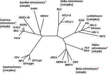
The family Retroviridae was previously divided into three subfamilies (Oncovirinae, Lentivirinae, and Spumavirinae), but are now divided into two: Orthoretrovirinae and Spumaretrovirinae. The term oncovirus is now commonly used to describe a cancer-causing virus. This family now includes the following genera:
- Subfamily Orthoretrovirinae:
- Genus Alpharetrovirus; type species: Avian leukosis virus; others include Rous sarcoma virus
- Genus Betaretrovirus; type species: Mouse mammary tumour virus
- Genus Gammaretrovirus; type species: Murine leukemia virus; others include Feline leukemia virus
- Genus Deltaretrovirus; type species: Bovine leukemia virus; others include the cancer-causing Human T-lymphotropic virus
- Genus Epsilonretrovirus; type species: Walleye dermal sarcoma virus
- Genus Lentivirus; type species: Human immunodeficiency virus 1; others include Simian, Feline immunodeficiency viruses
- Subfamily Spumaretrovirinae:
- Genus Bovispumavirus; type species: Bovine foamy virus
- Genus Equispumavirus; type species: Equine foamy virus
- Genus Felispumavirus; type species: Feline foamy virus
- Genus Prosimiispumavirus; type species: Brown greater galago prosimian foamy virus
- Genus Simiispumavirus; type species: Eastern chimpanzee simian foamy virus
Note that according to ICTV 2017, genus Spumavirus has been divided into five genera, and its former type species Simian foamy virus is now upgraded to genus Simiispumavirus with not less than 14 species, including new type species Eastern chimpanzee simian foamy virus.[16]
Group VII viruses
Both families in Group VII have DNA genomes contained within the invading virus particles. The DNA genome is transcribed into both mRNA, for use as a transcript in protein synthesis, and pre-genomic RNA, for use as the template during genome replication. Virally encoded reverse transcriptase uses the pre-genomic RNA as a template for the creation of genomic DNA.
Group VII includes:
- Family Caulimoviridae — e.g. Cauliflower mosaic virus
- Family Hepadnaviridae — e.g. Hepatitis B virus
The latter family is closely related to the newly proposed
- Family Nackednaviridae — e.g. African cichlid nackednavirus (ACNDV), formerly named African cichlid hepatitis B virus (ACHBV).[17]
whilst families Belpaoviridae, Metaviridae, Pseudoviridae, Retroviridae, and Caulimoviridae constitute the order Ortervirales.[18]
Endogenous
Endogenous retroviruses are not formally included in this classification system, and are broadly classified into three classes, on the basis of relatedness to exogenous genera:
- Class I are most similar to the gammaretroviruses
- Class II are most similar to the betaretroviruses and alpharetroviruses
- Class III are most similar to the spumaviruses.
Treatment
Antiretroviral drugs are medications for the treatment of infection by retroviruses, primarily HIV. Different classes of antiretroviral drugs act on different stages of the HIV life cycle. Combination of several (typically three or four) antiretroviral drugs is known as highly active anti-retroviral therapy (HAART).[19]
Treatment of veterinary retroviruses
Feline leukemia virus and Feline immunodeficiency virus infections are treated with biologics, including the only immunomodulator currently licensed for sale in the United States, Lymphocyte T-Cell Immune Modulator (LTCI).[20]
Notes
- The International Committee on Taxonomy of Viruses (ICTV) does not, however, consider retroviruses, which have DNA intermediates in their life cycle, as RNA viruses and classes them separately.[2]
References
- "Virus Taxonomy: 2018b Release". International Committee on Taxonomy of Viruses (ICTV). March 2019. Retrieved 16 March 2019.
- "Listing in Taxonomic Order – Index to ICTV Species Lists". Retrieved 11 April 2008.
- "retrovirus". Oxford English Dictionary. Retrieved 25 September 2018.
- Kurth, Reinhard; Bannert, Norbert, eds. (2010). Retroviruses: Molecular Biology, Genomics and Pathogenesis. Horizon Scientific. ISBN 978-1-904455-55-4.
- Coffin, John M. (1992). "Structure and Classification of Retroviruses". In Levy, Jay A. (ed.). The Retroviridae. 1 (1st ed.). New York: Plenum. p. 20. ISBN 978-0-306-44074-8.CS1 maint: ref=harv (link)
- Coffin 1992, pp. 26–34
- Kim FJ, Battini JL, Manel N, Sitbon M (January 2004). "Emergence of vertebrate retroviruses and envelope capture". Virology. 318 (1): 183–91. doi:10.1016/j.virol.2003.09.026. PMID 14972546.
- Vargiu, L; Rodriguez-Tomé, P; Sperber, GO; Cadeddu, M; Grandi, N; Blikstad, V; Tramontano, E; Blomberg, J (22 January 2016). "Classification and characterization of human endogenous retroviruses; mosaic forms are common". Retrovirology. 13: 7. doi:10.1186/s12977-015-0232-y. PMC 4724089. PMID 26800882.
- Coffin, John M.; Hughes, Stephen H.; Varmus, Harold E. (1997). "Genetic Organization". Retroviruses. Cold Spring Harbor Laboratory Press.
- Robert Belshaw; Pereira V; Katzourakis A; Talbot G; Paces J; Burt A; Tristem M. (April 2004). "Long-term reinfection of the human genome by endogenous retroviruses". Proc Natl Acad Sci USA. 101 (14): 4894–9. Bibcode:2004PNAS..101.4894B. doi:10.1073/pnas.0307800101. PMC 387345. PMID 15044706.
- Medstrand P, van de Lagemaat L, Dunn C, Landry J, Svenback D, Mager D (2005). "Impact of transposable elements on the evolution of mammalian gene regulation". Cytogenet Genome Res. 110 (1–4): 342–52. doi:10.1159/000084966. PMID 16093686.
- Svarovskaia ES; Cheslock SR; Zhang WH; Hu WS; Pathak VK. (January 2003). "Retroviral mutation rates and reverse transcriptase fidelity". Front. Biosci. 8 (1–3): d117–34. doi:10.2741/957. PMID 12456349.
- Jolly C (March 2011). "Cell-to-cell transmission of retroviruses: Innate immunity and interferon-induced restriction factors". Virology. 411 (2): 251–9. doi:10.1016/j.virol.2010.12.031. PMC 3053447. PMID 21247613.
- Aiewsakun, Pakorn; Katzourakis, Aris (2017). "Marine origin of retroviruses in the early Palaeozoic Era". Nature Communications. 8: 13954. Bibcode:2017NatCo...813954A. doi:10.1038/ncomms13954. PMC 5512871. PMID 28071651.
- Desport, M, ed. (2010). Lentiviruses and Macrophages: Molecular and Cellular Interactions. Caister Academic. ISBN 978-1-904455-60-8.
- ICTV Taxonomy Browser
- Lauber C, Seitz S, Mattei S, Suh A, Beck J, Herstein J, Börold J, Salzburger W, Kaderali L, Briggs JA, Bartenschlager R (September 2017). "Deciphering the Origin and Evolution of Hepatitis B Viruses by Means of a Family of Non-enveloped Fish Viruses". Cell Host & Microbe. 22 (3): 387–399.e6. doi:10.1016/j.chom.2017.07.019. PMC 5604429. PMID 28867387. and PDF
- Krupovic, M; Blomberg, J; Coffin, JM; Dasgupta, I; Fan, H; Geering, AD; Gifford, R; Harrach, B; Hull, R; Johnson, W; Kreuze, JF; Lindemann, D; Llorens, C; Lockhart, B; Mayer, J; Muller, E; Olszewski, N; Pappu, HR; Pooggin, M; Richert-Pöggeler, KR; Sabanadzovic, S; Sanfaçon, H; Schoelz, JE; Seal, S; Stavolone, L; Stoye, JP; Teycheney, PY; Tristem, M; Koonin, EV; Kuhn, JH (4 April 2018). "Ortervirales: A new viral order unifying five families of reverse-transcribing viruses". Journal of Virology. 92 (12). doi:10.1128/JVI.00515-18. PMC 5974489. PMID 29618642.
- Rutherford, G. W.; Sangani, P. R.; Kennedy, G. E. (2003). "Three- or four- versus two-drug antiretroviral maintenance regimens for HIV infection". The Cochrane Database of Systematic Reviews (4): CD002037. doi:10.1002/14651858.CD002037. ISSN 1469-493X. PMID 14583945.
- Gingerich, Daniel A. (2008). "Lymphocyte T-cell immunomodulator (LTCI): Review of the immunopharmacology of a new biologic" (PDF). International Journal of Applied Research in Veterinary Medicine. 6 (2): 61–68. ISSN 1559-470X.
External links
| Wikimedia Commons has media related to Retroviridae. |
- ViralZone A Swiss Institute of Bioinformatics resource for all viral families, providing general molecular and epidemiological information (follow links for "Retro-transcribing viruses")
- Retrovirus Animation (Flash Required)
- Retrovirology Scientific journal
- Retrovirus life cycle chapter From Kimball's Biology (online biology textbook pages)
- Coffin, John M; Hughes, Stephen H; Varmus, Harold E, eds. (1997). Retroviruses. Cold Spring Harbor Laboratory. ISBN 978-0-87969-571-2. NBK19376.
- Specter, Michael (3 December 2007). "Annals of Science: Darwin's Surprise". The New Yorker.
| Components |
|  |
|---|---|---|
| Viral life cycle |
| |
| Genetics |
| |
| By host |
| |
| Other |
| |
| ||
| Authority control |
|---|