Parietal eye
A parietal eye, also known as a third eye or pineal eye, is a part of the epithalamus present in some species of fish, amphibians and reptiles. The eye is located at the top of the head, is photoreceptive and is associated with the pineal gland, regulating circadian rhythmicity and hormone production for thermoregulation.[1]

Presence in various animals
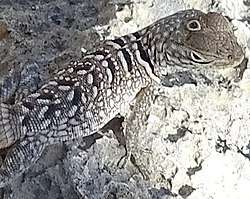
The parietal eye is found in the tuatara, most lizards, frogs, salamanders, certain bony fish, sharks, and lampreys (a kind of jawless fish).[2][3][4] It is absent in mammals, but was present in their closest extinct relatives, the therapsids.[5] It is also absent in turtles and archosaurs, which includes birds and crocodilians, and their extinct relatives.[6]
Anatomy
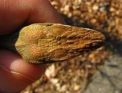
The third eye, where present, is always much smaller than the main paired eyes, and, in living species, it is always covered by skin, and is usually not readily visible externally.[7]
The parietal eye is a part of the epithalamus, which can be divided into two major parts; the epiphysis (the pineal organ, or pineal gland if mostly endocrine) and the parapineal organ (often called the parietal eye, or third eye if it is photoreceptive). The parietal eye arises as an anterior evagination of the pineal organ or as a separate outgrowth of the roof of the diencephalon. In some species, it protrudes through the skull.[8] The parietal eye uses a different biochemical method of detecting light from that of rod cells or cone cells in a normal vertebrate eye.[9]
Many of the oldest fossil vertebrates, including ostracoderms, placoderms, crossopterygians, and even early tetrapods, had a socket in the skull that appears to have held a functional third eye. This socket remains as a foramen between the parietal bones even in many living amphibians and reptiles, although it has vanished in birds and mammals.
Lampreys have two parietal eyes, one that developed from the parapineal organ and the other from the pineal organ. These are one behind the other in the centre of the upper surface of the braincase. Because lampreys are among the most primitive of all living vertebrates, it is possible that this was the original condition among vertebrates, and may have allowed bottom-dwelling species to sense threats from above.[7]
Saniwa, an extinct varanid lizard, probably had two parietal eyes, one that developed from the pineal organ and the other from the parapineal organ. Saniwa is the only known jawed vertebrate to have both a pineal and a parapineal eye. In most vertebrates, the pineal organ forms the parietal eye, however, in lepidosaurs, it is formed from the parapineal organ. This implies that Saniwa reevolved the pineal eye.[10]
Comparative anatomy
The parietal eye of amphibians and reptiles appears relatively far forward in the skull; thus it may be surprising that the human pineal gland appears far away from this position, tucked away between the corpus callosum and cerebellum. Also the parietal bones, in humans, make up a portion of the rear of the skull, far from the eyes. To understand this, note that the parietal bones formed a part of the skull lying between the eyes in sarcopterygians and basal amphibians, but have moved further back in higher vertebrates.[11] Likewise, in the brain of the frog, the diencephalon, from which the pineal stalk arises, appears relatively further forward, as the cerebral hemispheres are smaller but the optic lobes are far more prominent than the human mesencephalon, which is part of the brain stem.[12] In humans the optic tract, commissure, and optic nerve bridge the substantial distance between eyes and diencephalon. Likewise the pineal stalk of Petromyzon elongates very considerably during metamorphosis.[13]
Analogs in other species
Crustaceans have a single eye atop the head as a nauplius (first-stage larva). The eye has a lens and senses the direction of light but cannot resolve more details in images. Later, more sophisticated segmented eyes develop on sides of the head while the initial eye stays for some time. So, it is possible to say that, at some stage of development, crustaceans also have a "third eye". Some species, like the brine shrimp, retain the primary eye, being three-eyed in the adult stage. Most arthropods have simple eyes, called ocelli, between their main eyes.[14]
See also
- Third eye
- Arthropod eye
- Mollusc eye
- Simple eye in invertebrates
- Vision in fish
References
- Eakin, R. M (1973). The Third Eye. Berkeley: University of California Press.
- Dodt, Eberhard (1973). "The Parietal Eye (Pineal and Parietal Organs) of Lower Vertebrates". Visual Centers in the Brain. Handbook of Sensory Physiology. 7 / 3 / 3 B. Springer, Berlin, Heidelberg. pp. 113–140. doi:10.1007/978-3-642-65495-4_4. ISBN 9783642654978.
- Uetz, Peter (2003-10-07). "Sphenodontidae". The EMBL reptile database. European Molecular Biology Laboratory, heidelberg. Archived from the original on 2007-02-21. Retrieved 2007-02-22.
- Gundy, GC; Wurst, GZ (1976). "The occurrence of parietal eyes in recent Lacertilia (Reptilia)". Journal of Herpetology. 10 (2): 113–121. doi:10.2307/1562791. JSTOR 1562791.
- Benoit, Julien; Abdala, Fernando; Manger, Paul R.; Rubidge, Bruce S. (2016-03-17). "The Sixth Sense in Mammalian Forerunners: Variability of the Parietal Foramen and the Evolution of the Pineal Eye in South African Permo-Triassic Eutheriodont Therapsids". Acta Palaeontologica Polonica. 61 (4): 777–789. doi:10.4202/app.00219.2015. ISSN 0567-7920.
- Emerling, Christopher A. (2017-03-01). "Archelosaurian Color Vision, Parietal Eye Loss, and the Crocodylian Nocturnal Bottleneck". Molecular Biology and Evolution. 34 (3): 666–676. doi:10.1093/molbev/msw265. ISSN 1537-1719. PMID 27940498.
- Romer, Alfred Sherwood; Parsons, Thomas S. (1977). The Vertebrate Body. Philadelphia, PA: Holt-Saunders International. pp. 471–473. ISBN 978-0-03-910284-5.
- Zug, George; Vitt, Laurie Vitt; Caldwell, Janalee (2002). Herpetology: An Introductory Biology of Amphibians and Reptiles, Second Edition. San Diego: Academic Press. p. 75. ISBN 978-0-12-782622-6.
- Xiong, Wei-Hong; Solessio, Eduardo C.; Yau, King-Wai (1998). "An unusual cGMP pathway underlying depolarizing light response of the vertebrate parietal-eye photoreceptor". Nature Neuroscience. 1 (5): 359–65. doi:10.1038/1570. PMID 10196524. Retrieved 2007-02-22.
- Smith, Krister T.; Bhullar, Bhart-Anjan S.; Köhler, Gunther; Habersetzer, Jörg (2 April 2018). "The Only Known Jawed Vertebrate with Four Eyes and the Bauplan of the Pineal Complex". Current Biology. 28 (7): 1101–1107.e2. doi:10.1016/j.cub.2018.02.021. ISSN 0960-9822. PMID 29614279.
- "FRONTAL AND PARIETAL BONES=". Retrieved 2011-09-08.
- "Edible Frog Brain Clipart". Etc.usf.edu. Retrieved 2011-09-08.
- Journal of morphology - Google Books. 1887. Retrieved 2011-09-08.
- Mayer, Georg (2006-12-01). "Structure and development of onychophoran eyes: What is the ancestral visual organ in arthropods?". Arthropod Structure & Development. 35 (4): 231–245. doi:10.1016/j.asd.2006.06.003. ISSN 1467-8039. PMID 18089073.
Vision in animals | ||
|---|---|---|
| Vision | .jpg) | |
| Eyes |
| |
| Evolution | ||
| Coloration | ||
| Related topics | ||