Mite
| Mites | |
|---|---|
 | |
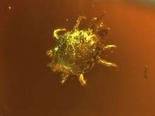
| Peacock mite (Tuckerella sp.), false color SEM, magnified 260× | |
| Scientific classification | |
| Kingdom: | Animalia |
| Phylum: | Arthropoda |
| Subphylum: | Chelicerata |
| Class: | Arachnida |
| Subclass: | Acari |
| Groups included | |
| Cladistically included but traditionally excluded taxa | |
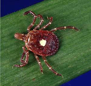
Mites are small arthropods belonging to the class Arachnida and the subclass Acari. Mites are not a clade as they span two different groups of arachnids: the Acariformes are sister to the camel spiders, while the Parasitiformes are sister to the false scorpions; also, they exclude the ticks, order Ixodida, although ticks and mites are closely related. Mites are distantly related to spiders and scorpions.
The body is in two sections, the cephalothorax or prosoma (there is no separate head), and an opisthosoma. The scientific discipline devoted to the study of ticks and mites is called acarology.
Most mites are tiny, less than 1 mm (0.04 in) in length, and have a simple, unsegmented body plan. Their small size makes them easily overlooked; some species live in water, many live in soil as decomposers, others live on plants, sometimes creating galls, while others again are predators or parasites. This last type includes the commercially important Varroa parasite of honey bees, as well as the scabies mite of humans. Most species are harmless to humans but a few are associated with allergies or may transmit diseases.
Evolution and taxonomy
_Lorryia_formosa_2_edit.jpg)
The mites are not an exact taxon, but the name is used for members of several groups in the subclass Acari. The phylogeny of the Acari has been relatively little studied, but molecular information from ribosomal DNA is being extensively used to understand relationships between groups. The 18 S rRNA gene provides information on relationships among phyla and superphyla, while the ITS2, and the 18S ribosomal RNA and 28S ribosomal RNA genes, provide clues at deeper levels.[1]
Taxonomy
The third edition (2009) of the standard textbook A Manual of Acarology uses a system of six orders, grouped into three superorders:[2]
- Superorder Opilioacariformes – a small order of large mites that superficially resemble harvestmen (Opiliones), hence their name
- Superorder Parasitiformes – ticks and a variety of mites
- Holothyrida - predatory mites from the southern hemisphere
- Ixodida – hard ticks and soft ticks
- Mesostigmata – bird mites, phytoseiid mites
- Trigynaspida - large, diverse order
- Monogynaspida - diverse order of parasitic and predatory mites
- Superorder Acariformes – the most diverse group of mites
- Trombidiformes – plant parasitic mites (spider mites, peacock mites, gall mites, red-legged earth mites, etc.), snout mites, chiggers, hair follicle mites, velvet mites, water mites, etc.
- Sphaerolichida - small order of mites containing two families
- Prostigmata - large order of sucking mites
- Sarcoptiformes
- Oribatida – oribatid mites, beetle mites, armored mites (also cryptostigmata)
- Astigmatina – stored product, fur, feather, dust, and human itch mites, etc.
- Trombidiformes – plant parasitic mites (spider mites, peacock mites, gall mites, red-legged earth mites, etc.), snout mites, chiggers, hair follicle mites, velvet mites, water mites, etc.
Fossil record
Most fossil acarids are no older than the Tertiary (up to 65 mya).[3] Earlier fossils are too few to enable mite phylogeny to be reconstructed from palaeontological evidence, but in 2002 an oribatid mite (Brachypylina) from the Early Ordovician (c. 480 mya) was found in Oland, Sweden.[4] The first find of Parasitiformes from the Mesozoic was of an argasid tick larva in Cretaceous amber (90–94 mya) from New Jersey.[5] Other fossils including the first opilioacariform mite are preserved in Baltic amber of Eocene age (44 mya).[6]
Phylogeny
Members of the superorders Opilioacariformes and Acariformes (sometimes known as Actinotrichida) are mites, as well as some of the Parasitiformes (sometimes known as Anactinotrichida).[7] Recent genetic research has caused a change in the naming scheme, however, and recent publications have changed the superorder Parasitiformes to an order.[8] Other recent research has suggested that Acari is polyphyletic (of multiple origins), with ticks and spiders more closely related than ticks and mites.[9] The cladogram is based on Dabert et al. 2010, which used molecular data. It shows the Acariformes sister to the Solifugae (camel spiders), while the Parasitiformes are sister to the Pseudoscorpionida.[10]
|
"Acari" (mites and ticks) |
.jpg)
.jpg)


Anatomy
External
Mites are tiny members of the class Arachnida; most are in the size range 250 to 750 μm (0.01 to 0.03 in) but some are larger and some are no bigger than 100 μm (0.004 in) as adults. The body plan is similar to that of ticks in having two regions, a cephalothorax (with no separate head) or prosoma, and an opisthosoma or abdomen. Segmentation has almost entirely been lost and the prosoma and opisthosoma are fused, only the positioning of the limbs indicating the location of the segments.[11]
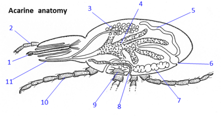
At the front of the body is the gnathosoma or capitulum. This is not a head and does not contain the eyes or the brain, but is a retractable feeding apparatus consisting of the chelicerae, the pedipalps and the oral cavity. It is covered above by an extension of the body carapace and is connected to the body by a flexible section of cuticle. The mouthparts differ between taxa depending on diet; in some species the appendages resemble legs while in others they are modified into chelicerae-like structures. The oral cavity connects posteriorly to the mouth and pharynx.[11]
Most mites have four pairs of legs, each with six segments, which may be modified for swimming or other purposes. The dorsal surface of the body is clad in hardened tergites and the ventral surface by hardened sclerites; sometimes these form transverse ridges. The gonopore (genital opening) is located on the ventral surface between the fourth pair of legs. Some species have one to five median or lateral eyes but many species are blind, and slit and pit sense organs are common. Both body and limbs bear setae (bristles) which may be simple, flattened, club-shaped or sensory. Mites are usually some shade of brown, but some species are red, orange, black or green, or some combination of these colours.[11]
Internal
Mites have a typical arachnid digestive system, although some species lack an anus: they do not defecate during their short lives.[13] The circulatory system consists of a network of sinuses and lacks a heart, movement of fluid being driven by the contraction of body muscles. Gas exchange is carried out across the body surface, but many species additionally have between one and four pairs of tracheae, the spiracles being located in the front half of the body. The excretory system includes a nephridium and one or two pairs of Malpighian tubules.[11]
Reproduction and life cycle
The sexes are separate in mites; males have a pair of testes in the mid-region of the body, each connected to the gonopore by a vas deferens, and in some species there is a chitinous penis; females have a single ovary connected to the gonopore by an oviduct, as well as a seminal receptacle for the storage of sperm. In most mites, sperm is transferred to the female indirectly; the male either deposits a spermatophore on a surface from which it is picked up by the female, or he uses his chelicerae or third pair of legs to insert it into the female's gonopore. In some of the Acariformes, insemination is direct using the male's penis.[11]
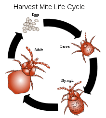
The eggs are laid in the substrate, or wherever the mite happens to live. They take up to six weeks to hatch, according to species, and the first stage larvae have six legs. After three moults, the larvae become nymphs, with eight legs, and after a further three moults, they become adults. Longevity varies between species, but the lifespan of mites is short as compared to many other arachnids.[11]
Ecology
Niches
Mites occupy a wide range of ecological niches. For example, Oribatida mites are important decomposers in many habitats. They eat a wide variety of material including living and dead plant and fungal material, lichens and carrion; some are predatory, though no oribatid mites are parasitic.[15] Mites are among the most diverse and successful of all invertebrate groups. They have exploited a wide array of habitats, and because of their small size go largely unnoticed. They are found in fresh and salt water, in the soil, in forests, pastures, agricultural crops, ornamental plants, thermal springs and caves. They inhabit organic debris of all kinds and are extremely numerous in leaf litter. They feed on animals, plants and fungi and some are parasites of plants and animals.[16] Some 48,200 species of mites have been described,[17] but there may be a million or more species as yet undescribed.[11] The tropical species Archegozetes longisetosus is one of the strongest animals in the world, relative to its mass (100 μg): It lifts up to 1,182 times its own weight, over five times more than would be expected of such a minute animal.[18] A mite also holds a speed record: for its length, Paratarsotomus macropalpis is the fastest animal on Earth.[19][20]
Parasitism
Many mites are parasitic on plants and animals. One family of mites, Pyroglyphidae, or nest mites, live primarily in the nests of birds and animals. These mites are largely parasitic and consume blood, skin and keratin. Dust mites, which feed mostly on dead skin and hair shed from humans instead of consuming them from the organism directly, evolved from these parasitic ancestors.[21]
Parasitic mites sometimes infest insects. Varroa destructor attaches to the body of honey bees, and Acarapis woodi (family Tarsonemidae) lives in their tracheae. Hundreds of species are associated with other bees, mostly poorly described. They attach to bees in a variety of ways. For example, Trigona corvina workers have been found with mites attached to the outer face of their hind tibiae.[22] Some are thought to be parasites, while others are beneficial symbionts. Mites also parasitize some ant species, such as Eciton burchellii.[23]
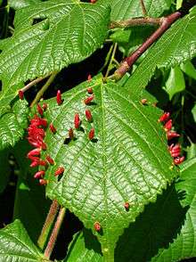
Plant pests include the so-called spider mites (family Tetranychidae), thread-footed mites (family Tarsonemidae), and the gall mites (family Eriophyidae).[24] Among the species that attack animals are members of the sarcoptic mange mites (family Sarcoptidae), which burrow under the skin. Demodex mites (family Demodicidae) are parasites that live in or near the hair follicles of mammals, including humans.[25]
Dispersal
Being unable to fly, mites need some other means of dispersal. On a small scale, walking is used to access other suitable locations in the immediate vicinity. Some species mount to a high point and adopt a dispersal posture and get carried away by the wind, while others waft a thread of silk aloft to balloon to a new position.[26]
Parasitic mites use their hosts to disperse, and spread from host to host by direct contact. Another strategy is phoresy; the mite, often equipped with suitable claspers or suckers, grips onto an insect or other animal, and gets transported to another place. A phoretic mite is just a hitch-hiker and does not feed during the time it is carried by its temporary host. These travelling mites are mostly species that reproduce rapidly and are quick to colonise new habitats.[26]
Relationship with humans

Mites are tiny, almost invisible, and apart from those that are of economic concern to humans, little studied. The majority are beneficial, living in the soil or aqueous environments and assisting in the decomposition of decaying organic material, or consuming fungi, plant or animal matter, as part of the carbon cycle.[16]
Medical significance
The majority of mite species are harmless to humans and domestic animals, but a few species can colonize mammals directly, acting as vectors for disease transmission, and causing or contributing to allergenic diseases. Mites which colonize human skin are the cause of several types of itchy skin rashes, such as gamasoidosis,[27] rodent mite dermatitis,[28] grain itch,[29] grocer's itch,[29] and scabies; Sarcoptes scabiei is a parasitic mite responsible for scabies, which is one of the three most common skin disorders in children.[30] Demodex mites, which are common cause of mange in dogs and other domesticated animals,[25] have also been implicated in the human skin disease rosacea, although the mechanism by which demodex contributes to the disease is unclear.[31]

Chiggers are known primarily for their itchy bite, but they can also spread disease in some limited circumstances, such as scrub typhus.[32] The house-mouse mite is the only known vector of the disease rickettsialpox.[33] House dust mites, found in warm and humid places such as beds, cause several forms of allergic diseases, including hay fever, asthma and eczema, and are known to aggravate atopic dermatitis.[34]
Among domestic animals, sheep are affected by the mite Psoroptes ovis which lives on the skin, causing hypersensitivity and inflammation.[35]
In beekeeping
The mite Varroa destructor is a serious pest of honey bees, contributing to colony collapse disorder in commercial hives. The mite is an obligate external parasite, able to reproduce only in bee colonies. It directly weakens its host by sucking up the bee's fat, and can spread RNA viruses including deformed wing virus. Heavy infestation causes the death of a colony, generally over the winter. Since 2006, over 10 million beehives have been lost.[36][37]
In culture
Mites were first observed under the microscope by the English polymath Robert Hooke. In his 1665 book Micrographia, he stated that far from being spontaneously generated from dirt, they were "very prettily shap'd Insects".[38] The world's first science documentary featured cheese mites, seen under the microscope; the short film was shown in London's Alhambra music hall in 1903, causing a boom in the sales of simple microscopes.[38] A few years later, Arthur Conan Doyle wrote a satirical poem, Parable, with the conceit of some cheese mites disputing the origin of the round Cheddar cheese in which they all lived.[38]
See also
References
- Dhooria, Manjit Singh (2016). Fundamentals of Applied Acarology. Springer. p. 176. ISBN 978-981-10-1594-6.
- Gerald W. Krantz; D. E. Walter, eds. (2009). A Manual of Acarology (3rd ed.). Texas Tech University Press. ISBN 978-0-89672-620-8.
- de la Fuente, José (2003). "The fossil record and the origin of ticks (Acari: Parasitiformes: Ixodida)". Experimental and Applied Acarology. 29 (3/4): 331–344. doi:10.1023/A:1025824702816. PMID 14635818.
- Bernini, F.; Carnevale, G.; Bagnoli, G.; Stouge, S. (2002). "An Early Ordovician oribatid mite (Acari: Oribatida) from the island of Oland, Sweden". In Bernini, F.; Nannelli, R.; Nuzaci, G.; de Lillo, E. (eds.). Acarid Phylogeny and Evolution: Adaptation in Mites and Ticks. Proceedings of the IV Symposium of the European Association of Acarologists. Springer. pp. 45–47. ISBN 978-94-017-0611-7.
- Klompen, Hans; Grimaldi, David (2001). "First Mesozoic Record of a Parasitiform Mite: a Larval Argasid Tick in Cretaceous Amber (Acari: Ixodida: Argasidae)". Annals of the Entomological Society of America. 94 (1): 10–15. doi:10.1603/0013-8746(2001)094[0010:FMROAP]2.0.CO;2.
- Dunlop, Jason A.; Wunderlich, Jörg; Poinar, George O. (2007). "The first fossil opilioacariform mite (Acari: Opilioacariformes) and the first Baltic amber camel spider (Solifugae)". Transactions of the Royal Society of Edinburgh: Earth Sciences. 94 (3): 261–273. doi:10.1017/S0263593300000663.
- Walter, David Evans; Krantz, Gerald; Lindquist, Evert (13 December 1996). "Acari: The mites". Tree of Life Web Project. Retrieved 6 October 2017.
- Barker, S.C. & Murrell, A. (2004). "Systematics and evolution of ticks with a list of valid genus and species names". Parasitology. 129 (7): S15–S36. doi:10.1017/S0031182004005207. PMID 15938503.
- Sanggaard, Kristian W.; Bechsgaard, Jesper S.; Fang, Xiaodong (6 May 2014). "Spider genomes provide insight into composition and evolution of venom and silk". Nature Communications. 5: 3765. Bibcode:2014NatCo...5.3765S. doi:10.1038/ncomms4765. PMC 4273655. PMID 24801114.
- Dabert, Miroslawa; Witalinski, Wojciech; Kazmierski, Andrzej; Olszanowski, Ziemowit; Dabert, Jacek (2010). "Molecular phylogeny of acariform mites (Acari, Arachnida): Strong conflict between phylogenetic signal and long-branch attraction artifacts". Molecular Phylogenetics and Evolution. 56 (1): 222–241. doi:10.1016/j.ympev.2009.12.020. PMID 20060051.
- Ruppert, Edward E.; Fox, Richard, S.; Barnes, Robert D. (2004). Invertebrate Zoology, 7th edition. Cengage Learning. pp. 590–595. ISBN 978-81-315-0104-7.
- Balashov, Y.S. (1972). "Bloodsucking Ticks - Vectors of Diseases of Man and Animals". Miscellaneous Publications of the Entomological Society of America. 8: 161–376.
- Yong, Ed (27 August 2014). "You Almost Certainly Have Mites On Your Face". National Geographic. Retrieved 23 November 2017.
- Biljana D. Magud; Ljubiša Ž. Stanisavljević; Radmila U. Petanović (2007). "Morphological variation in different populations of Aceria anthocoptes (Acari: Eriophyoidea) associated with the Canada thistle, Cirsium arvense, in Serbia". Experimental and Applied Acarology. 42 (3): 173–183. doi:10.1007/s10493-007-9085-y. PMID 17611806.
- Arroyo, J.; Keith, A.M.; Schmidt, O.; Bolger, T. (2013). "Mite abundance and richness in an Irish survey of soil biodiversith with comments on some newly recorded species". Ir. Nat. J. 33: 19–27.
- Jeppson, L.R.; Keifer, Hartford H.; Baker, Edward William (1975). Mites Injurious to Economic Plants. University of California Press. pp. 1–3. ISBN 978-0-520-02381-9.
- R. B. Halliday, B. M. OConnor & A. S. Baker (2000). "Global Diversity of Mites". In Peter H. Raven; Tania Williams (eds.). Nature and human society: the quest for a sustainable world : proceedings of the 1997 Forum on Biodiversity. National Academies. pp. 192–212.
- Michael Heethoff & Lars Koerner (2007). "Small but powerful – the oribatid mite Archegozetes longisetosus Aoki (Acari, Oribatida) produces disproportionate high forces". Journal of Experimental Biology. 210 (17): 3036–3042. doi:10.1242/jeb.008276. PMID 17704078.
- Federation of American Societies for Experimental Biology (FASEB) (27 April 2014). "Mite sets new record as world's fastest land animal". Featured Research. ScienceDaily. Retrieved 23 November 2017.
- Rubin, Samuel; Young, Maria Ho-Yan; Wright, Jonathan C.; Whitaker, Dwight L.; Ahn, Anna N. (2016). "Exceptional running and turning performance in a mite". Journal of Experimental Biology. 219 (5): 676–685. doi:10.1242/jeb.128652. PMID 26787481.
- Erickson, Jim (2013-03-08). "Genetic study of house dust mites demonstrates reversible evolution". Michigan News. Retrieved 31 May 2014.
- Schwarz, Herbert Ferlando; Louise, Bacon, Annette (1948). "Stingless bees (Meliponidae) of the Western Hemisphere : Lestrimelitta and the following subgenera of Trigona : Trigona, Paratrigona, Schwarziana, Parapartamona, Cephalotrigona, Oxytrigona, Scaura, and Mourella. Bulletin of the AMNH ; v. 90". hdl:2246/1231. Cite journal requires
|journal=(help) - Berghoff, S.M.; Wurst, E.; Ebermann, E.; Sendova-Franks, A.A.B.; Rettenmeyer, C.W.; Franks, N.R. (2009). "Symbionts of societies that fission: Mites as guests or parasites of army ants". Ecological Entomology. 34 (6): 684–695. doi:10.1111/j.1365-2311.2009.01125.x.
- Fenemore, P.G. (2016). Plant Pests and Their Control. Elsevier. p. 112. ISBN 978-1-4831-8286-5.
- Hall, John C.; Hall, Brian J. (2009). Skin Infections: Diagnosis and Treatment. Cambridge University Press. p. 260. ISBN 978-0-521-89729-7.
- Capinera, John L. (2008). Encyclopedia of Entomology. Springer Science & Business Media. p. 2425. ISBN 978-1-4020-6242-1.
- Schulze, Keith E.; Cohen, Philip R. (1994). "Dove-associated gamasoidosis: A case of avian mite dermatitis". Journal of the American Academy of Dermatology. 30 (2): 278–280. doi:10.1016/S0190-9622(08)81930-5. ISSN 0190-9622. PMID 8288795.
- Theis, Jerold (1981-06-01). "Tropical Rat Mite Dermatitis". Archives of Dermatology. 117 (6): 341–3. doi:10.1001/archderm.1981.01650060031018. ISSN 0003-987X. PMID 7247425.
- James, William D.; Berger, Timothy G. (2006). Andrews' Diseases of the Skin: Clinical Dermatology. Saunders Elsevier. p. 454. ISBN 978-0-7216-2921-6.
- Andrews RM, McCarthy J, Carapetis JR, Currie BJ (December 2009). "Skin disorders, including pyoderma, scabies, and tinea infections". Pediatr. Clin. North Am. 56 (6): 1421–40. doi:10.1016/j.pcl.2009.09.002. PMID 19962029.
- Whitehead, Joanne; Barrows, Brady (2010). Journal of the Rosacea Research & Development Institute: Volume 1 Number 1, 2010. Universe. p. 47. ISBN 978-1-4502-0344-9.
- Pham, X.D.; Otsuka, Y.; Suzuki, H.; Takaoka, H. (2001). "Detection of Orientia tsutsugamushi (Rickettsiales: Rickettsiaceae) in unengorged chiggers (Acari: Trombiculidae) from Oita Prefecture, Japan, by nested polymerase chain reaction". Journal of Medical Entomology. 38 (2): 308–311. doi:10.1603/0022-2585-38.2.308. PMID 11296840.
- Diaz, J. H. (2010). "Endemic mite-transmitted dermatoses and infectious diseases in the South". The Journal of the Louisiana State Medical Society. 162 (3): 140–145, 147–149. PMID 20666166.
- Paul Klenerman; Brian Lipworth. "House dust mite allergy". NetDoctor. Retrieved February 20, 2008.
- Van den Broek, A. (2000). "Cutaneous and systemic responses during primary and challenge infestations of sheep with the sheep scab mite, Psoroptes ovis". Parasite Immunology. 22 (8): 407–414. doi:10.1046/j.1365-3024.2000.00318.x. PMID 10972847.
- Ernesto Guzmán-Novoa; Leslie Eccles; Yireli Calvete; Janine Mcgowan; Paul G. Kelly & Adriana Correa-Benítez (2009). "Varroa destructor is the main culprit for the death and reduced populations of overwintered honey bee (Apis mellifera) colonies in Ontario, Canada" (PDF). Apidologie. 41 (4): 443–450. doi:10.1051/apido/2009076.
- Benjamin, Alison (2 May 2010). "Fears for crops as shock figures from America show scale of bee catastrophe". The Guardian. London.
- Marren, Peter; Mabey, Richard (2010). Bugs Britannica. Chatto & Windus. pp. 122–125. ISBN 978-0-7011-8180-2.
External links
- Bitingmites.org: What's biting you?
- Worldwide honey bee decline due to mite infestations – article, photographs
- Mites and Ticks chapter in United States Environmental Protection Agency and University of Florida/Institute of Food and Agricultural Sciences National Public Health Pesticide Applicator Training Manual
- Mites at the US National Library of Medicine Medical Subject Headings (MeSH)
| Acariformes |
|  | |||||||
|---|---|---|---|---|---|---|---|---|---|
| Parasitiformes |
| ||||||||