Lamina affixa
Lamina affixa is a layer of epithelium growing on the surface of the thalamus and forming the floor of the central part of lateral ventricle, on whose medial margin is attached the choroid plexus of the lateral ventricle; it covers the superior thalamostriate vein and the superior choroidal vein. The torn edge of this plexus is called the tela choroidea.[1]
| Lamina affixa | |
|---|---|
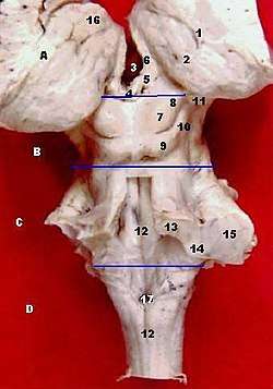
| |
 Human brain left dissected midsagittal view (Lamina affixa is #10) | |
| Details | |
| Identifiers | |
| Latin | lamina affixa |
| TA | A14.1.09.276 |
| FMA | 83709 |
| Anatomical terms of neuroanatomy | |
On the surface of the terminal vein is a narrow white band, named the lamina affixa.
References
This article incorporates text in the public domain from page 838 of the 20th edition of Gray's Anatomy (1918)
- Alberts, Daniel; et al. (2012). Dorland's illustrated medical dictionary (32nd ed.). Philadelphia, PA: Saunders/Elsevier. p. 1878. ISBN 978-1-4160-6257-8.
- Schober A, Böttner M, Strelau J, et al. (October 2001). "Expression of growth differentiation factor-15/ macrophage inhibitory cytokine-1 (GDF-15/MIC-1) in the perinatal, adult, and injured rat brain". J. Comp. Neurol. 439 (1): 32–45. doi:10.1002/cne.1333. PMID 11579380.
External links
| Authority control |
|---|
This article is issued from Wikipedia. The text is licensed under Creative Commons - Attribution - Sharealike. Additional terms may apply for the media files.