Fibrinoid necrosis
Fibrinoid necrosis is a specific pattern of irreversible, uncontrolled cell death that occurs when antigen-antibody complexes are deposited in the walls of blood vessels along with fibrin. It is common in the immune-mediated vasculitides which are a result of type III hypersensitivity. When stained with hematoxylin and eosin, they appear brightly eosinophilic and smudged.[1]
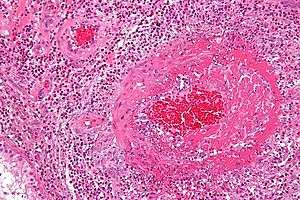
Micrograph showing (intensely pink) fibrinoid necrosis (large blood vessel - right of image) in a case of vasculitis (eosinophilic granulomatosis with polyangiitis). H&E stain.
Diseases
Fibrinoid necrosis is not limited to the immune-mediated vasculitides; many pathologic processes can lead to areas of fibrinoid necrosis. In systemic lupus erythematosus, the dermis is often affected by fluid accumulation and inflammation around the small vessels in the skin, which may show prominent fibrinoid necrosis.[1]
References
- Robbins and Cotran pathologic basis of disease. Kumar, Vinay, Abbas, Abul K., Aster, Jon C., Perkins, James A. (Ninth ed.). Philadelphia, PA. 2014. ISBN 9781455726134. OCLC 879416939.CS1 maint: others (link)
This article is issued from Wikipedia. The text is licensed under Creative Commons - Attribution - Sharealike. Additional terms may apply for the media files.