Coagulative necrosis
Coagulative necrosis is a type of accidental cell death typically caused by ischemia or infarction. In coagulative necrosis the architectures of dead tissue is preserved for at least a couple of days.[1] It is believed that the injury denatures structural proteins as well as lysosomal enzymes thus blocking the proteolysis of the damaged cells. The lack of lysosomal enzymes allows it to maintain a "coagulated" morphology for some time. Like most types of necrosis if enough viable cells are present around the affected area regeneration will usually occur.
Coagulative necrosis can also be induced by high local temperature; it is a desired effect of treatments such as high intensity focused ultrasound applied to cancerous cells.[2]
Causes
Coagulative necrosis is most commonly caused by conditions that do not involve severe trauma, toxins or an acute or chronic immune response. The lack of oxygen (hypoxia) causes cell death in a localised area which is perfused by blood vessels failing to deliver primarily oxygen, but also other important nutrients. It is important to note that while ischemia in most tissues of the body will cause coagulative necrosis, in the central nervous system ischemia causes liquefactive necrosis, as there is very little structural framework in neural tissue.
Pathology
Macroscopic
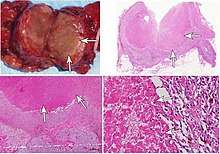
The macroscopic appearance of an area of coagulative necrosis is a pale segment of tissue contrasting against surrounding well vascularised tissue and is dry on cut surface. The tissue may later turn red due to inflammatory response. The surrounding surviving cells can aid in regeneration of the affected tissue unless they are stable or permanent.
Regeneration
As the majority of the structural remnants of the necrotic tissue remains, labile cells adjacent to the affected tissue will replicate and replace the cells which have been killed during the event. Labile cells are constantly undergoing mitosis and can therefore help reform the tissue, whereas nearby stable and permanent cells (e.g. neurons and cardiomyocytes) do not undergo mitosis and will not replace the tissue affected. Fibroblasts will also migrate to the affected area depositing fibrous tissue producing fibrosis or scarring in areas where viable cells do not replicate and replace tissue.
References
- Robbins and Cotran: Pathologic Basis of Disease, 8th Ed. 2010. Pg. 15
- Wu, F; Wang, Z-B; Cao, Y-De; Chen, W-Z; Bai, J; Zou, J-Z; Zhu, H (2003). "A randomised clinical trial of high-intensity focused ultrasound ablation for the treatment of patients with localised breast cancer". British Journal of Cancer. 89 (12): 2227–2233. doi:10.1038/sj.bjc.6601411. ISSN 0007-0920.
- Page 4 in: Clara Milikowski (1997). Color Atlas of Basic Histopathology. McGraw-Hill Professional Publishing. ISBN 978-0-8385-1382-8.