Squid giant synapse
The squid giant synapse is a chemical synapse found in squid. It is the largest chemical junction in nature.
Anatomy
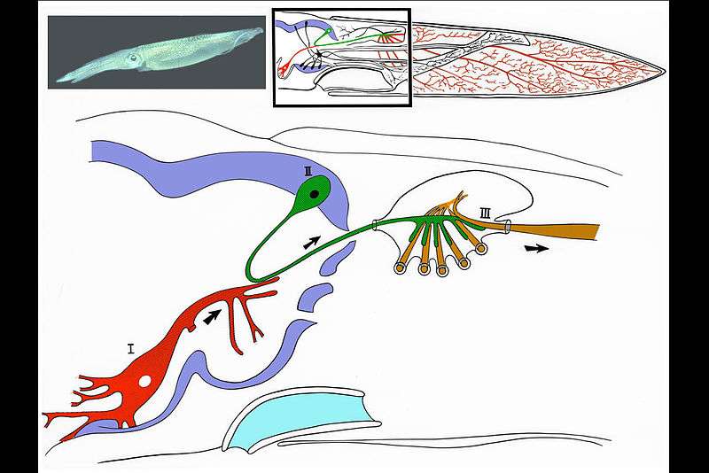
The squid giant synapse (Fig 1) was first recognized by John Zachary Young in 1939. It lies in the stellate ganglion on each side of the midline, at the posterior wall of the squid’s muscular mantle. Activation of this synapse triggers a synchronous contraction of the mantle musculature, causing the forceful ejection of a jet of water from the mantle. This water propulsion allows the squid to move rapidly through the water and even to jump through the surface of the water (breaking the air-water barrier) to escape predators.
The signal to the mantle is transmitted via a chain consisting of three giant neurons organized in sequence. The first is located in the ventral magnocellular lobe, central to the eyes. It serves as a central integrating manifold that receives all sensory systems and consists of two symmetrical neurons (I). They, in turn, contact secondary neurons (one in each side) in the dorsal magnocellular lobe and (II) and in turn contact the tertiary giant axons in the stellate ganglion (III, one in each side of the mantle). These latter are the giant axons that the work of Alan Hodgkin and Andrew Huxley made famous. Each secondary axon branches at the stellate ganglion and contacts all the tertiary axons; thus, information concerning relevant sensory input is relayed from the sense organs in the cephalic ganglion (the squid’s brain) to the contractile muscular mantle (which is activated directly by the tertiary giant axons).
Electrophysiology
Many essential elements of how all chemical synapses function were first discovered by studying the squid giant synapse. Early electrophysiological studies demonstrated the chemical nature of transmission at this synapse by making simultaneous intracellular recording from the presynaptic and postsynaptic terminals in vitro (Bullock & Hagiwara 1957, Hagiwara & Tasaki 1958, Takeuchi & Takeuchi 1962). Classical experiments later on demonstrated that, in the absence of action potentials, transmission could occur (Bloedel et al. 1966, Katz & Miledi 1967, Kusano, Livengood & Werman 1967). The calcium hypothesis for synaptic transmission was directly demonstrated in this synapse by showing that at the equilibrium potential for calcium, no transmitter is released (Katz & Miledi 1967). Thus, calcium entry and not the change in the transmembrane electric field per se is responsible for transmitter release (Llinás et al. 1981, Augustine, Charlton & Smith 1985). This preparation continues to be the most useful for the study of the molecular and cell biological basis for transmitter release. Other important new mammalian preparations are now available for such studies such as the calyx of Held.

See also
References
- Young, John Z. (25 May 1939). "Fused neurons and synaptic contacts in the giant nerve fibers of cephalopods". Philosophical Transactions of the Royal Society B: Biological Sciences. 229 (564): 465–503. doi:10.1098/rstb.1939.0003 – via The Royal Society.
- Bullock, Theodore H.; Hagiwara, Susumu (20 March 1957). "Intracellular recording from the giant synapse of the squid". Journal of General Physiology. 40 (4): 565–577. doi:10.1085/jgp.40.4.565 – via Rockefeller University Press.
- Hagiwara, S.; Tasaki, I. (29 August 1958). "A study on the mechanism of impulse transmission across the giant synapse of the squid". Journal of Physiology. 143 (1): 114–137. doi:10.1113/jphysiol.1958.sp006048. PMC 1356715. PMID 13576464 – via Wiley Online Library.
- Takeuchi, Akira; Takeuchi, Noriko (July 1962). "Electrical changes in the pre and postsynaptic axons of the giant synapse of Loligo". Journal of General Physiology. 45 (6): 1181–1193. doi:10.1085/jgp.45.6.1181. PMC 2195243. PMID 13919241 – via Rockefeller University Press.
- Bloedel, J.R.; Gage, P.W.Q.; Llinás, R.; Quastel, D.M.J. (October 1966). "Transmitter release at the squid giant synapse in the presence of tetrodotoxin". Nature. 212: 49–50. doi:10.1038/212049a0. PMID 5965569 – via Nature Publishing Group.
- Katz, B.; Miledi, R. (September 1967). "A study of synaptic transmission in the absence of nerve impulses". Journal of Physiology. 192 (2): 407–436. doi:10.1113/jphysiol.1967.sp008307. PMC 1365564. PMID 4383089 – via Wiley Online Library.
- Kusano, Kiyoshi; Livengood, David R.; Werman, Robert (December 1967). "Correlation of transmitter release with membrane properties of the presynaptic fiber of the squid giant synapse". Journal of General Physiology. 50 (11): 2579–2601. doi:10.1085/jgp.50.11.2579. PMC 2225670. PMID 4296572 – via Rockefeller University Press.
- Llinás, Rodolfo; Steinberg, Izchak Z.; Walton, Kerry (August 1976). "Presynaptic calcium currents and their relation to synaptic transmission. Voltage clamp study in the squid giant synapse and theoretical model for the calcium gate" (PDF). Proceedings of the National Academy of Sciences of the United States of America. 73 (8): 2918–2922. doi:10.1073/pnas.73.8.2918. PMC 430802. PMID 183215 – via PNAS.
- Llinás, R.; Steinberg, I.Z.; Walton, K. (March 1981a). "Presynaptic calcium currents in the squid giant synapse". Biophysical Journal. 33 (3): 289–321. doi:10.1016/S0006-3495(81)84898-9 – via Cell Press.
- Llinás, R.; Steinberg, I.Z.; Walton, K. (March 1981b). "Relationship between presynaptic calcium current and postsynaptic potential in squid giant synapse". Biophysical Journal. 33 (3): 323–352. doi:10.1016/S0006-3495(81)84899-0 – via Cell Press.
- Augustine, G.J.; Charlton, M.P.; Smith, S.J. (October 1985). "Calcium entry into voltage clamped presynaptic terminals of squid". J. Physiol. 367 (1): 163–181. doi:10.1113/jphysiol.1985.sp015819. PMC 1193058. PMID 2865362 – via Wiley Online Library.
- Llinás, Rodolfo R. (1999). The squid Giant Synapse. New York: Oxford University Press. ISBN 0-19-511652-6. OCLC 934117310.
Cephalopod anatomy | |||||||||
|---|---|---|---|---|---|---|---|---|---|
| Shell |
|    | |||||||
| Mantle and funnel |
| ||||||||
| Head and limbs |
| ||||||||
| General |
| ||||||||
Developmental stages: Spawn → Paralarva (Doratopsis stage) → Juvenile → Subadult → Adult • Egg fossils • Protoconch (embryonic shell) | |||||||||