Preload (cardiology)
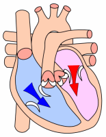
In cardiac physiology, preload is the end diastolic volume that stretches the right or left ventricle of the heart to its greatest dimensions under variable physiologic demand.[1] In other words, it is the initial stretching of the cardiomyocytes prior to contraction; therefore, it is related to the sarcomere length at the end of diastole. Parameters such as ventricular end diastolic volume or pressure are used to measure preload since the ideal length of the cardiac sarcomere cannot be measured. Passive filling of the (heart) ventricle and subsequent atrial contraction thus allows an echocardiographically volumetric measurement. Preload is theoretically most accurately described as the initial stretching of a single cardiomyocyte prior to contraction. This cannot be measured in vivo and therefore other measurements are used as estimates. Estimation may be inaccurate, for example in a chronically dilated ventricle new sarcomeres may have formed in the heart muscle allowing the relaxed ventricle to appear enlarged. The term end-diastolic volume is better suited to the clinic, although not exactly equivalent to the strict definition of preload. Atrial pressure is a surrogate for preload.
Calculation
Quantitatively, preload can be calculated as

where:
LVEDP = Left ventricular end diastolic pressure
LVEDR = Left ventricular end diastolic radius (at the ventricle's midpoint)
h = thickness of the ventricle.
This calculation is based on the Law of Laplace which states that (wall stress) = . Hence, preload is the wall stress. Preload is measured in pressure units (mm Hg).

Factors affecting preload
Preload is affected by venous blood pressure and the rate of venous return. These are affected by venous tone and volume of circulating blood.
Preload is related to the ventricular end-diastolic volume; a higher end-diastolic volume implies a higher preload. However, the relationship is not simple because of the restriction of the term preload to single myocytes. Preload can still be approximated by the inexpensive echocardiographic measurement end diastolic volume or EDV.
Preload increases with exercise (slightly), increasing blood volume (overtransfusion, polycythemia) and neuroendocrine excitement (sympathetic tone).
An arteriovenous fistula can increase preload.[2]
Preload is also affected by two main body "pumps":
Respiratory pump - Intrapleural pressure decreases during inspiration and abdominal pressure increases, squeezing local abdominal veins, allowing thoracic veins to expand and increase blood flow towards the right atrium.
Skeletal muscle pump - In the deep veins of the legs, surrounding muscles squeeze veins and pump blood back towards the heart. This occurs most notably in the legs. Once blood flows past valves it cannot flow backwards and therefore blood is “milked” towards the heart.
See also
References
- ↑ Leucke, thomas; Roth, Harry (1993). "Assessment of cardiac preload and left ventricular function under increasing levels of positive end-expiratory pressure". Intensive Care Medicine. 30 (1): 119–126. doi:10.1007/s00134-003-1993-7. Retrieved 13 July 2011.
- ↑ "Pulmonary: Heart Failure". Archived from the original on 2009-02-01. Retrieved 2008-12-21.