Posterior inferior cerebellar artery
| Posterior inferior cerebellar artery | |
|---|---|
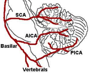 | |
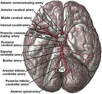
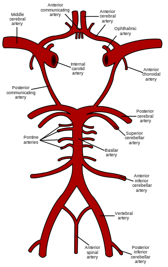 Diagram of the arterial circulation at the base of the brain (PICA labeled at bottom right) | |
| Details | |
| Source | Vertebral artery |
| Branches |
Medial branch lateral |
| Vein | Inferior cerebellar veins |
| Supplies | Cerebellum, choroid plexus of the fourth ventricle |
| Identifiers | |
| Latin | Arteria cerebelli inferior posterior |
| TA | A12.2.08.012 |
| FMA | 50518 |
|
Anatomical terminology | |
The posterior inferior cerebellar artery (PICA), the largest branch of the vertebral artery, is one of the three main arterial blood supplies for the cerebellum, part of the brain. Occlusion of the posterior inferior cerebellar artery or one of its branches, or of the vertebral artery leads to lateral medullary syndrome also called Wallenberg syndrome.
Course
It winds backward around the upper part of the medulla oblongata, passing between the origins of the vagus nerve and the accessory nerve, over the inferior cerebellar peduncle to the undersurface of the cerebellum, where it divides into two branches.
The medial branch continues backward to the notch between the two hemispheres of the cerebellum; while the lateral supplies the under surface of the cerebellum, as far as its lateral border, where it anastomoses with the anterior inferior cerebellar and the superior cerebellar branches of the basilar artery.
Branches from this artery supply the choroid plexus of the fourth ventricle.
Clinical significance
A disrupted blood supply to this artery due to a blood clot or embolus will result in tissue damage and lead to lateral medullary syndrome, also known as posterior inferior cerebellar artery syndrome (PICA syndrome), or Wallenberg syndrome. Severe occlusion of this artery or to vertebral arteries could lead to Horner's Syndrome as well. Lateral medullary syndrome has many neurological symptoms.
References
This article incorporates text in the public domain from page 580 of the 20th edition of Gray's Anatomy (1918)
External links
- Anatomy photo:28:09-0225 at the SUNY Downstate Medical Center
- "Anatomy diagram: 13048.000-1". Roche Lexicon - illustrated navigator. Elsevier. Archived from the original on 2014-01-01.