Perilymph
| Perilymph | |
|---|---|
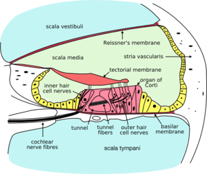 Cross-section of cochlea. Perilymph is located in the scala vestibuli and scala tympani - the aqua regions at the top and bottom of the diagram. | |
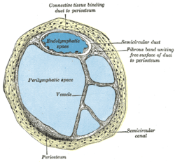 Cross-section of semi-circular canal and duct showing perilymphatic space | |
| Details | |
| Identifiers | |
| Latin | perilympha |
| MeSH | D010498 |
| TA | A15.3.03.056 |
| FMA | 60908 |
| Anatomical terminology | |
Perilymph is an extracellular fluid located within the inner ear. It is found within the semicircular canals and the scala tympani and scala vestibuli of the cochlea. The ionic composition of perilymph is comparable to that of plasma and cerebrospinal fluid. The major cation in perilymph is sodium, with the values of sodium and potassium concentration in the perilymph being 138 mM and 6.9 mM, respectively.[1] It is also named Cotunnius' liquid and liquor cotunnii for Domenico Cotugno.
Structure
The inner ear has two parts: the bony labyrinth and the membranous labyrinth. The membranous labyrinth is contained within the bony labyrinth, and contains a fluid called endolymph. Between the outer wall of the membranous labyrinth and the wall of the bony labyrinth is the perilymphatic space which contains the perilymph. The membranous labyrinth is suspended in the perilymph. The perilymph in the bony labyrinth is continuous with the cerebrospinal fluid of the subarachnoid space via the perilymphatic duct.[2]
Composition
Perilymph and endolymph have unique ionic compositions suited to their functions in regulating electrochemical impulses of hair cells. The electric potential of endolymph is ~80-90 mV more positive than perilymph due to a higher concentration of potassium cations (K+) than sodium (Na+).[3]
Perilymph is the fluid contained within the bony labyrinth, surrounding and protecting the membranous labyrinth; perilymph resembles extracellular fluid in composition (sodium salts are the predominate positive electrolyte) and, via the cochlear aqueduct (sometimes referred to as the "perilymphatic duct"), is in continuity with cerebrospinal fluid.
Endolymph is the fluid contained within the membranous labyrinth of the inner ear; endolymph resembles intracellular fluid in composition (potassium is the main cation).
Clinical significance
It has also been suggested that perilymph and endolymph participate in a unidirectional flow that is interrupted in Ménière's disease.
References
- ↑ Bosher SK, Warren RL (1968-11-05). "Observations on the electrochemistry of the cochlear endolymph of the rat: a quantitative study of its electrical potential and ionic composition as determined by means of flame spectrophotometry". Proceedings of the Royal Society B. 171 (1023): 227–247. doi:10.1098/rspb.1968.0066. PMID 4386844.
- ↑ Blumenfeld, Hal (2010). Neuroanatomy through Clinical Cases second edition. Sinauer Associates, Inc.
- ↑ Konishi T, Hamrick PE, Walsh PJ (1978). "Ion transport in guinea pig cochlea. I. Potassium and sodium transport". Acta Otolaryngol. 86 (1–2): 22–34. doi:10.3109/00016487809124717. PMID 696294.