Pericardial sinus
| Pericardial sinus | |
|---|---|
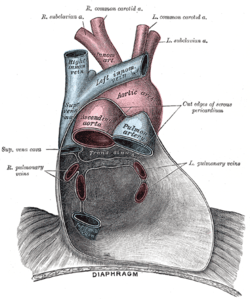 Posterior wall of the pericardial sac, showing the lines of reflection of the serous pericardium on the great vessels. (Transverse sinus labeled at center. Oblique sinus not labeled, but visible inferior to transverse sinus between the right and left pulmonary veins) | |
| Identifiers | |
| TA | A12.1.07.001 |
| FMA | 77132 |
| Anatomical terminology | |
There are two pericardial sinuses: transverse and oblique.
- The cul-de-sac sinus, enclosed between the limbs of the inverted U of the venous mesocardium lies posterior to the left atrium and is known as the oblique sinus.
- The passage between the venous and arterial mesocardia—i.e., between the aorta and pulmonary artery anteriorly and the superior vena cava posteriorly —is termed the transverse sinus.[1] Also, the sinus that forms in the pericardial cavity where the dorso-mesentary pericardium reside. Note: This sinus is clinically important because passing one end of clamp through the sinus, and the other end anterior to the aorta/pulmonary trunk will allow complete blockage of blood output. This is done during some heart surgeries.
- Can be used to pass ligature during cardiac surgery.
References
This article incorporates text in the public domain from page 526 of the 20th edition of Gray's Anatomy (1918)
External links
- Anatomy photo:20:04-0101 at the SUNY Downstate Medical Center - "Heart: Transverse and Oblique Pericardial Sinuses"
- thoraxlesson4 at The Anatomy Lesson by Wesley Norman (Georgetown University) (pericardialsinuses)
This article is issued from
Wikipedia.
The text is licensed under Creative Commons - Attribution - Sharealike.
Additional terms may apply for the media files.