PSMD1

26S proteasome non-ATPase regulatory subunit 1, also as known as 26S Proteasome Regulatory Subunit Rpn2 (systematic nomenclature), is a protein that in humans is encoded by the PSMD1 gene.[5][6] This protein is one of the 19 essential subunits that contributes to the complete assembly of 19S proteasome complex.[7]
Structure
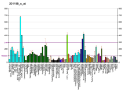
Gene expression
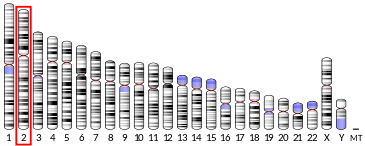
The gene PSMD1 encodes the largest non-ATPase subunit of the 19S regulator base, which is responsible for substrate recognition and binding.[6] The human PSMD1 gene has 25 exons and locates at chromosome band 2q37.1. The human protein 26S proteasome non-ATPase regulatory subunit 1 is 106 kDa in size and composed of 953 amino acids. The calculated theoretical pI of this protein is 5.25. An alternative splicing during gene expression generates an isoform of the protein in which the amino acid sequence from 797-827 is missing.
Complex assembly
26S proteasome complex is usually consisted of a 20S core particle (CP, or 20S proteasome) and one or two 19S regulatory particles (RP, or 19S proteasome) on either one side or both side of the barrel-shaped 20S. The CP and RPs pertain distinct structural characteristics and biological functions. In brief, 20S sub complex presents three types proteolytic activities, including caspase-like, trypsin-like, and chymotrypsin-like activities. These proteolytic active sites located in the inner side of a chamber formed by 4 stacked rings of 20S subunits, preventing random protein-enzyme encounter and uncontrolled protein degradation. The 19S regulatory particles can recognize ubiquitin-labeled protein as degradation substrate, unfold the protein to linear, open the gate of 20S core particle, and guide the substate into the proteolytic chamber. To meet such functional complexity, 19S regulatory particle contains at least 18 constitutive subunits. These subunits can be categorized into two classes based on the ATP dependence of subunits, ATP-dependent subunits and ATP-independent subunits. According to the protein interaction and topological characteristics of this multisubunit complex, the 19S regulatory particle is composed of a base and a lid subcomplex. The base consists of a ring of six AAA ATPases (Subunit Rpt1-6, systematic nomenclature) and four non-ATPase subunits (Rpn1, Rpn2, Rpn10, and Rpn13). Protein 26S proteasome non-ATPase regulatory subunit 1 (Rpn2) is an essential component of forming the base sub complex of 19S regulatory particle. Traditionally, Rpn1 and Rpn2 were considered residing at the center of base sub complex and surrounded by six AAA ATPases (Rpt 1-6). However, recent investigation provides an alternative structure of 19S base via an integrative approach combining data from cryoelectron microscopy, X-ray crystallography, residue-specific chemical cross-linking, and several proteomics techniques. Rpn2 is rigid protein located on the side of ATPase ring, supporting as the connection between the lid and base. Rpn1 is conformationally variable, positioned at the periphery of the ATPase ring. The ubiquitin receptors Rpn10 and Rpn13 are located further in the distal part of the 19S complex, indicating that they were recruited to the complex late during the assembly process.[8]
Function
As the degradation machinery that is responsible for ~70% of intracellular proteolysis,[9] proteasome complex (26S proteasome) plays a critical roles in maintaining the homeostasis of cellular proteome. Accordingly, misfolded proteins and damaged protein need to be continuously removed to recycle amino acids for new synthesis; in parallel, some key regulatory proteins fulfill their biological functions via selective degradation; furthermore, proteins are digested into peptides for MHC class I antigen presentation. To meet such complicated demands in biological process via spatial and temporal proteolysis, protein substrates have to be recognized, recruited, and eventually hydrolyzed in a well controlled fashion. Thus, 19S regulatory particle pertains a series of important capabilities to address these functional challenges. To recognize protein as designated substrate, 19S complex has subunits that are capable to recognize proteins with a special degradative tag, the ubiquitinylation. It also have subunits that can bind with nucleotides (e.g., ATPs) in order to facilitate the association between 19S and 20S particles, as well as to cause confirmation changes of alpha subunit C-terminals that form the substate entrance of 20S complex. Rpn2 is the largest subunit of 19S regulatory particle and stays at the center of the "base" subcomplex of 19S particle.
Clinical significance
The Proteasome and its subunits are of clinical significance for at least two reasons: (1) a compromised complex assembly or a dysfunctional proteasome can be associated with the underlying pathophysiology of specific diseases, and (2) they can be exploited as drug targets for therapeutic interventions. More recently, more effort has been made to consider the proteasome for the development of novel diagnostic markers and strategies. An improved and comprehensive understanding of the pathophysiology of the proteasome should lead to clinical applications in the future.
The proteasomes form a pivotal component for the Ubiquitin-Proteasome System (UPS) [10] and corresponding cellular Protein Quality Control (PQC). Protein ubiquitination and subsequent proteolysis and degradation by the proteasome are important mechanisms in the regulation of the cell cycle, cell growth and differentiation, gene transcription, signal transduction and apoptosis.[11] Subsequently, a compromised proteasome complex assembly and function lead to reduced proteolytic activities and the accumulation of damaged or misfolded protein species. Such protein accumulation may contribute to the pathogenesis and phenotypic characteristics in neurodegenerative diseases,[12][13] cardiovascular diseases,[14][15][16] inflammatory responses and autoimmune diseases,[17] and systemic DNA damage responses leading to malignancies.[18]
Several experimental and clinical studies have indicated that aberrations and deregulations of the UPS contribute to the pathogenesis of several neurodegenerative and myodegenerative disorders, including Alzheimer's disease,[19] Parkinson's disease[20] and Pick's disease,[21] Amyotrophic lateral sclerosis (ALS),[21] Huntington's disease,[20] Creutzfeldt–Jakob disease,[22] and motor neuron diseases, polyglutamine (PolyQ) diseases, Muscular dystrophies[23] and several rare forms of neurodegenerative diseases associated with dementia.[24] As part of the Ubiquitin-Proteasome System (UPS), the proteasome maintains cardiac protein homeostasis and thus plays a significant role in cardiac Ischemic injury,[25] ventricular hypertrophy[26] and Heart failure.[27] Additionally, evidence is accumulating that the UPS plays an essential role in malignant transformation. UPS proteolysis plays a major role in responses of cancer cells to stimulatory signals that are critical for the development of cancer. Accordingly, gene expression by degradation of transcription factors, such as p53, c-Jun, c-Fos, NF-κB, c-Myc, HIF-1α, MATα2, STAT3, sterol-regulated element-binding proteins and androgen receptors are all controlled by the UPS and thus involved in the development of various malignancies.[28] Moreover, the UPS regulates the degradation of tumor suppressor gene products such as adenomatous polyposis coli (APC) in colorectal cancer, retinoblastoma (Rb). and von Hippel-Lindau tumor suppressor (VHL), as well as a number of proto-oncogenes (Raf, Myc, Myb, Rel, Src, Mos, Abl). The UPS is also involved in the regulation of inflammatory responses. This activity is usually attributed to the role of proteasomes in the activation of NF-κB which further regulates the expression of pro inflammatory cytokines such as TNF-α, IL-β, IL-8, adhesion molecules (ICAM-1, VCAM-1, P-selectin) and prostaglandins and nitric oxide (NO)[17] Additionally, the UPS also plays a role in inflammatory responses as regulators of leukocyte proliferation, mainly through proteolysis of cyclines and the degradation of CDK inhibitors.[29] Lastly, autoimmune disease patients with SLE, Sjogren's syndrome and rheumatoid arthritis (RA) predominantly exhibit circulating proteasomes which can be applied as clinical biomarkers.[30]
A clinical study on patients with age related macula degeneration identified four significant proteins, including 26S proteasome non-ATPase regulatory subunit 1 (Rpn2), that were increased, according to semi-quantitative proteomic profiling. The study reported that an LC-MRM assay revealed a significant increase of Rpn2 in 15 macula degeneration patients compared to the control subjects, suggesting that this protein could be a biomarker for this condition.[31] Age-related macular degeneration is the leading cause of blindness in the world. Evidence is accumulating that the suppression of the UPS contributes to the increase of toxic proteins and inflammation in retina pigment epithelium, the functional abnormalities and/or the degeneration of which are believed to be the initiators and major pathologies of macula degeneration.[32] There are only limited options for the treatment of macular degeneration, thus early identification of susceptibility and preventive measures are important therapeutic strategies. New potential biomarkers for neovascular macular degeneratuon and UPS-related proteins that are altered in patients such as Rpn2 may serve as the basis for future clinical studies to determine target proteins involved in the protection of the eye against macula degeneration.[31][32]
References
- 1 2 3 GRCh38: Ensembl release 89: ENSG00000173692 - Ensembl, May 2017
- 1 2 3 GRCm38: Ensembl release 89: ENSMUSG00000026229 - Ensembl, May 2017
- ↑ "Human PubMed Reference:".
- ↑ "Mouse PubMed Reference:".
- ↑ Yokota K, Kagawa S, Shimizu Y, Akioka H, Tsurumi C, Noda C, Fujimuro M, Yokosawa H, Fujiwara T, Takahashi E, Ohba M, Yamasaki M, DeMartino GN, Slaughter CA, Toh-e A, Tanaka K (Jun 1996). "CDNA cloning of p112, the largest regulatory subunit of the human 26s proteasome, and functional analysis of its yeast homologue, sen3p". Molecular Biology of the Cell. 7 (6): 853–70. doi:10.1091/mbc.7.6.853. PMC 275938. PMID 8816993.
- 1 2 "Entrez Gene: PSMD1 proteasome (prosome, macropain) 26S subunit, non-ATPase, 1".
- ↑ Gu ZC, Enenkel C (Dec 2014). "Proteasome assembly". Cellular and Molecular Life Sciences. 71 (24): 4729–45. doi:10.1007/s00018-014-1699-8. PMID 25107634.
- ↑ Lasker K, Förster F, Bohn S, Walzthoeni T, Villa E, Unverdorben P, Beck F, Aebersold R, Sali A, Baumeister W (Jan 2012). "Molecular architecture of the 26S proteasome holocomplex determined by an integrative approach". Proceedings of the National Academy of Sciences of the United States of America. 109 (5): 1380–7. Bibcode:2012PNAS..109.1380L. doi:10.1073/pnas.1120559109. PMC 3277140. PMID 22307589.
- ↑ Rock KL, Gramm C, Rothstein L, Clark K, Stein R, Dick L, Hwang D, Goldberg AL (Sep 1994). "Inhibitors of the proteasome block the degradation of most cell proteins and the generation of peptides presented on MHC class I molecules". Cell. 78 (5): 761–71. doi:10.1016/s0092-8674(94)90462-6. PMID 8087844.
- ↑ Kleiger G, Mayor T (Jun 2014). "Perilous journey: a tour of the ubiquitin-proteasome system". Trends in Cell Biology. 24 (6): 352–9. doi:10.1016/j.tcb.2013.12.003. PMC 4037451. PMID 24457024.
- ↑ Goldberg AL, Stein R, Adams J (Aug 1995). "New insights into proteasome function: from archaebacteria to drug development". Chemistry & Biology. 2 (8): 503–8. doi:10.1016/1074-5521(95)90182-5. PMID 9383453.
- ↑ Sulistio YA, Heese K (Jan 2015). "The Ubiquitin-Proteasome System and Molecular Chaperone Deregulation in Alzheimer's Disease". Molecular Neurobiology. 53: 905–31. doi:10.1007/s12035-014-9063-4. PMID 25561438.
- ↑ Ortega Z, Lucas JJ (2014). "Ubiquitin-proteasome system involvement in Huntington's disease". Frontiers in Molecular Neuroscience. 7: 77. doi:10.3389/fnmol.2014.00077. PMC 4179678. PMID 25324717.
- ↑ Sandri M, Robbins J (Jun 2014). "Proteotoxicity: an underappreciated pathology in cardiac disease". Journal of Molecular and Cellular Cardiology. 71: 3–10. doi:10.1016/j.yjmcc.2013.12.015. PMC 4011959. PMID 24380730.
- ↑ Drews O, Taegtmeyer H (Dec 2014). "Targeting the ubiquitin-proteasome system in heart disease: the basis for new therapeutic strategies". Antioxidants & Redox Signaling. 21 (17): 2322–43. doi:10.1089/ars.2013.5823. PMC 4241867. PMID 25133688.
- ↑ Wang ZV, Hill JA (Feb 2015). "Protein quality control and metabolism: bidirectional control in the heart". Cell Metabolism. 21 (2): 215–26. doi:10.1016/j.cmet.2015.01.016. PMC 4317573. PMID 25651176.
- 1 2 Karin M, Delhase M (Feb 2000). "The I kappa B kinase (IKK) and NF-kappa B: key elements of proinflammatory signalling". Seminars in Immunology. 12 (1): 85–98. doi:10.1006/smim.2000.0210. PMID 10723801.
- ↑ Ermolaeva MA, Dakhovnik A, Schumacher B (Sep 2015). "Quality control mechanisms in cellular and systemic DNA damage responses". Ageing Research Reviews. 23 (Pt A): 3–11. doi:10.1016/j.arr.2014.12.009. PMC 4886828. PMID 25560147.
- ↑ Checler F, da Costa CA, Ancolio K, Chevallier N, Lopez-Perez E, Marambaud P (Jul 2000). "Role of the proteasome in Alzheimer's disease". Biochimica et Biophysica Acta. 1502 (1): 133–8. doi:10.1016/s0925-4439(00)00039-9. PMID 10899438.
- 1 2 Chung KK, Dawson VL, Dawson TM (Nov 2001). "The role of the ubiquitin-proteasomal pathway in Parkinson's disease and other neurodegenerative disorders". Trends in Neurosciences. 24 (11 Suppl): S7–14. doi:10.1016/s0166-2236(00)01998-6. PMID 11881748.
- 1 2 Ikeda K, Akiyama H, Arai T, Ueno H, Tsuchiya K, Kosaka K (Jul 2002). "Morphometrical reappraisal of motor neuron system of Pick's disease and amyotrophic lateral sclerosis with dementia". Acta Neuropathologica. 104 (1): 21–8. doi:10.1007/s00401-001-0513-5. PMID 12070660.
- ↑ Manaka H, Kato T, Kurita K, Katagiri T, Shikama Y, Kujirai K, Kawanami T, Suzuki Y, Nihei K, Sasaki H (May 1992). "Marked increase in cerebrospinal fluid ubiquitin in Creutzfeldt–Jakob disease". Neuroscience Letters. 139 (1): 47–9. doi:10.1016/0304-3940(92)90854-z. PMID 1328965.
- ↑ Mathews KD, Moore SA (Jan 2003). "Limb-girdle muscular dystrophy". Current Neurology and Neuroscience Reports. 3 (1): 78–85. doi:10.1007/s11910-003-0042-9. PMID 12507416.
- ↑ Mayer RJ (Mar 2003). "From neurodegeneration to neurohomeostasis: the role of ubiquitin". Drug News & Perspectives. 16 (2): 103–8. doi:10.1358/dnp.2003.16.2.829327. PMID 12792671.
- ↑ Calise J, Powell SR (Feb 2013). "The ubiquitin proteasome system and myocardial ischemia". American Journal of Physiology. Heart and Circulatory Physiology. 304 (3): H337–49. doi:10.1152/ajpheart.00604.2012. PMC 3774499. PMID 23220331.
- ↑ Predmore JM, Wang P, Davis F, Bartolone S, Westfall MV, Dyke DB, Pagani F, Powell SR, Day SM (Mar 2010). "Ubiquitin proteasome dysfunction in human hypertrophic and dilated cardiomyopathies". Circulation. 121 (8): 997–1004. doi:10.1161/CIRCULATIONAHA.109.904557. PMC 2857348. PMID 20159828.
- ↑ Powell SR (Jul 2006). "The ubiquitin-proteasome system in cardiac physiology and pathology". American Journal of Physiology. Heart and Circulatory Physiology. 291 (1): H1–H19. doi:10.1152/ajpheart.00062.2006. PMID 16501026.
- ↑ Adams J (Apr 2003). "Potential for proteasome inhibition in the treatment of cancer". Drug Discovery Today. 8 (7): 307–15. doi:10.1016/s1359-6446(03)02647-3. PMID 12654543.
- ↑ Ben-Neriah Y (Jan 2002). "Regulatory functions of ubiquitination in the immune system". Nature Immunology. 3 (1): 20–6. doi:10.1038/ni0102-20. PMID 11753406.
- ↑ Egerer K, Kuckelkorn U, Rudolph PE, Rückert JC, Dörner T, Burmester GR, Kloetzel PM, Feist E (Oct 2002). "Circulating proteasomes are markers of cell damage and immunologic activity in autoimmune diseases". The Journal of Rheumatology. 29 (10): 2045–52. PMID 12375310.
- 1 2 Lee H, Choi AJ, Kang GY, Park HS, Kim HC, Lim HJ, Chung H (May 2014). "Increased 26S proteasome non-ATPase regulatory subunit 1 in the aqueous humor of patients with age-related macular degeneration". BMB Reports. 47 (5): 292–7. doi:10.5483/bmbrep.2014.47.5.193. PMC 4163863. PMID 24286321.
- 1 2 Pickart CM (2001). "Mechanisms underlying ubiquitination". Annual Review of Biochemistry. 70: 503–33. doi:10.1146/annurev.biochem.70.1.503. PMID 11395416.
Further reading
- Coux O, Tanaka K, Goldberg AL (1996). "Structure and functions of the 20S and 26S proteasomes". Annual Review of Biochemistry. 65: 801–47. doi:10.1146/annurev.bi.65.070196.004101. PMID 8811196.
- Goff SP (Aug 2003). "Death by deamination: a novel host restriction system for HIV-1". Cell. 114 (3): 281–3. doi:10.1016/S0092-8674(03)00602-0. PMID 12914693.
- Seeger M, Ferrell K, Frank R, Dubiel W (Mar 1997). "HIV-1 tat inhibits the 20 S proteasome and its 11 S regulator-mediated activation". The Journal of Biological Chemistry. 272 (13): 8145–8. doi:10.1074/jbc.272.13.8145. PMID 9079628.
- Madani N, Kabat D (Dec 1998). "An endogenous inhibitor of human immunodeficiency virus in human lymphocytes is overcome by the viral Vif protein". Journal of Virology. 72 (12): 10251–5. PMC 110608. PMID 9811770.
- Simon JH, Gaddis NC, Fouchier RA, Malim MH (Dec 1998). "Evidence for a newly discovered cellular anti-HIV-1 phenotype". Nature Medicine. 4 (12): 1397–400. doi:10.1038/3987. PMID 9846577.
- Lüders J, Demand J, Höhfeld J (Feb 2000). "The ubiquitin-related BAG-1 provides a link between the molecular chaperones Hsc70/Hsp70 and the proteasome". The Journal of Biological Chemistry. 275 (7): 4613–7. doi:10.1074/jbc.275.7.4613. PMID 10671488.
- Mulder LC, Muesing MA (Sep 2000). "Degradation of HIV-1 integrase by the N-end rule pathway". The Journal of Biological Chemistry. 275 (38): 29749–53. doi:10.1074/jbc.M004670200. PMID 10893419.
- Sheehy AM, Gaddis NC, Choi JD, Malim MH (Aug 2002). "Isolation of a human gene that inhibits HIV-1 infection and is suppressed by the viral Vif protein". Nature. 418 (6898): 646–50. Bibcode:2002Natur.418..646S. doi:10.1038/nature00939. PMID 12167863.
- Huang X, Seifert U, Salzmann U, Henklein P, Preissner R, Henke W, Sijts AJ, Kloetzel PM, Dubiel W (Nov 2002). "The RTP site shared by the HIV-1 Tat protein and the 11S regulator subunit alpha is crucial for their effects on proteasome function including antigen processing". Journal of Molecular Biology. 323 (4): 771–82. doi:10.1016/S0022-2836(02)00998-1. PMID 12419264.
- Gaddis NC, Chertova E, Sheehy AM, Henderson LE, Malim MH (May 2003). "Comprehensive investigation of the molecular defect in vif-deficient human immunodeficiency virus type 1 virions". Journal of Virology. 77 (10): 5810–20. doi:10.1128/JVI.77.10.5810-5820.2003. PMC 154025. PMID 12719574.
- Lecossier D, Bouchonnet F, Clavel F, Hance AJ (May 2003). "Hypermutation of HIV-1 DNA in the absence of the Vif protein". Science. 300 (5622): 1112. doi:10.1126/science.1083338. PMID 12750511.
- Zhang H, Yang B, Pomerantz RJ, Zhang C, Arunachalam SC, Gao L (Jul 2003). "The cytidine deaminase CEM15 induces hypermutation in newly synthesized HIV-1 DNA". Nature. 424 (6944): 94–8. Bibcode:2003Natur.424...94Z. doi:10.1038/nature01707. PMC 1350966. PMID 12808465.
- Mangeat B, Turelli P, Caron G, Friedli M, Perrin L, Trono D (Jul 2003). "Broad antiretroviral defence by human APOBEC3G through lethal editing of nascent reverse transcripts". Nature. 424 (6944): 99–103. Bibcode:2003Natur.424...99M. doi:10.1038/nature01709. PMID 12808466.
- Harris RS, Bishop KN, Sheehy AM, Craig HM, Petersen-Mahrt SK, Watt IN, Neuberger MS, Malim MH (Jun 2003). "DNA deamination mediates innate immunity to retroviral infection". Cell. 113 (6): 803–9. doi:10.1016/S0092-8674(03)00423-9. PMID 12809610.
- Harris RS, Sheehy AM, Craig HM, Malim MH, Neuberger MS (Jul 2003). "DNA deamination: not just a trigger for antibody diversification but also a mechanism for defense against retroviruses". Nature Immunology. 4 (7): 641–3. doi:10.1038/ni0703-641. PMID 12830140.
- Gu Y, Sundquist WI (Jul 2003). "Good to CU". Nature. 424 (6944): 21–2. Bibcode:2003Natur.424...21G. doi:10.1038/424021a. PMID 12840737.
- Mariani R, Chen D, Schröfelbauer B, Navarro F, König R, Bollman B, Münk C, Nymark-McMahon H, Landau NR (Jul 2003). "Species-specific exclusion of APOBEC3G from HIV-1 virions by Vif". Cell. 114 (1): 21–31. doi:10.1016/S0092-8674(03)00515-4. PMID 12859895.