Polyadenylate-binding protein 4 (PABPC4) is a protein that in humans is encoded by the PABPC4 gene.[5][6]
Function
Poly(A)-binding proteins (PABPs) bind to the poly(A) tail present at the 3-prime ends of most eukaryotic mRNAs. PABPC4 or IPABP (inducible PABP) was isolated as an activation-induced T-cell mRNA encoding a protein. Activation of T cells increased PABPC4 mRNA levels in T cells approximately 5-fold. PABPC4 contains 4 RNA-binding domains and proline-rich C terminus. PABPC4 is localized primarily to the cytoplasm. It is suggested that PABPC4 might be necessary for regulation of stability of labile mRNA species in activated T cells. PABPC4 was also identified as an antigen, APP1 (activated-platelet protein-1), expressed on thrombin-activated rabbit platelets. PABPC4 may also be involved in the regulation of protein translation in platelets and megakaryocytes or may participate in the binding or stabilization of polyadenylates in platelet dense granules.[6]
References
- 1 2 3 GRCh38: Ensembl release 89: ENSG00000090621 - Ensembl, May 2017
- 1 2 3 GRCm38: Ensembl release 89: ENSMUSG00000011257 - Ensembl, May 2017
- ↑ "Human PubMed Reference:".
- ↑ "Mouse PubMed Reference:".
- ↑ Féral C, Mattéi MG, Pawlak A, Guellaën G (Nov 1999). "Chromosomal localization of three human poly(A)-binding protein genes and four related pseudogenes". Hum Genet. 105 (4): 347–53. doi:10.1007/s004390051113. PMC 1865476. PMID 10543404.
- 1 2 "Entrez Gene: PABPC4 poly(A) binding protein, cytoplasmic 4 (inducible form)".
- ↑ "Glucose tolerance test data for Pabpc4". Wellcome Trust Sanger Institute.
- 1 2 3 Gerdin, AK (2010). "The Sanger Mouse Genetics Programme: High throughput characterisation of knockout mice". Acta Ophthalmologica. 88: 925–7. doi:10.1111/j.1755-3768.2010.4142.x.
- ↑ Mouse Resources Portal, Wellcome Trust Sanger Institute.
- ↑ "International Knockout Mouse Consortium".
- ↑ "Mouse Genome Informatics".
- ↑ Skarnes WC, Rosen B, West AP, Koutsourakis M, Bushell W, Iyer V, Mujica AO, Thomas M, Harrow J, Cox T, Jackson D, Severin J, Biggs P, Fu J, Nefedov M, de Jong PJ, Stewart AF, Bradley A (2011). "A conditional knockout resource for the genome-wide study of mouse gene function". Nature. 474 (7351): 337–342. doi:10.1038/nature10163. PMC 3572410. PMID 21677750.
- ↑ Dolgin E (2011). "Mouse library set to be knockout". Nature. 474 (7351): 262–3. doi:10.1038/474262a. PMID 21677718.
- ↑ Collins FS, Rossant J, Wurst W (2007). "A Mouse for All Reasons". Cell. 128 (1): 9–13. doi:10.1016/j.cell.2006.12.018. PMID 17218247.
- ↑ van der Weyden L, White JK, Adams DJ, Logan DW (2011). "The mouse genetics toolkit: revealing function and mechanism". Genome Biol. 12 (6): 224. doi:10.1186/gb-2011-12-6-224. PMC 3218837. PMID 21722353.
- ↑ Hinz T, Flindt S, Marx A, Janssen O, Kabelitz D (May 2001). "Inhibition of protein synthesis by the T cell receptor-inducible human TDAG51 gene product". Cell. Signal. 13 (5): 345–52. doi:10.1016/S0898-6568(01)00141-3. PMID 11369516.
Further reading
- Maruyama K, Sugano S (1994). "Oligo-capping: a simple method to replace the cap structure of eukaryotic mRNAs with oligoribonucleotides". Gene. 138 (1–2): 171–4. doi:10.1016/0378-1119(94)90802-8. PMID 8125298.
- Yang H, Duckett CS, Lindsten T (1996). "iPABP, an inducible poly(A)-binding protein detected in activated human T cells". Mol. Cell. Biol. 15 (12): 6770–6. PMC 230930. PMID 8524242.
- Houng AK, Maggini L, Clement CY, Reed GL (1997). "Identification and structure of activated-platelet protein-1, a protein with RNA-binding domain motifs that is expressed by activated platelets". Eur. J. Biochem. 243 (1–2): 209–18. doi:10.1111/j.1432-1033.1997.0209a.x. PMID 9030741.
- Suzuki Y, Yoshitomo-Nakagawa K, Maruyama K, Suyama A, Sugano S (1997). "Construction and characterization of a full length-enriched and a 5'-end-enriched cDNA library". Gene. 200 (1–2): 149–56. doi:10.1016/S0378-1119(97)00411-3. PMID 9373149.
- Hoshino S, Imai M, Kobayashi T, Uchida N, Katada T (1999). "The eukaryotic polypeptide chain releasing factor (eRF3/GSPT) carrying the translation termination signal to the 3'-Poly(A) tail of mRNA. Direct association of erf3/GSPT with polyadenylate-binding protein". J. Biol. Chem. 274 (24): 16677–80. doi:10.1074/jbc.274.24.16677. PMID 10358005.
- Hinz T, Flindt S, Marx A, Janssen O, Kabelitz D (2001). "Inhibition of protein synthesis by the T cell receptor-inducible human TDAG51 gene product". Cell. Signal. 13 (5): 345–52. doi:10.1016/S0898-6568(01)00141-3. PMID 11369516.
- Li J, Hawkins IC, Harvey CD, Jennings JL, Link AJ, Patton JG (2003). "Regulation of Alternative Splicing by SRrp86 and Its Interacting Proteins". Mol. Cell. Biol. 23 (21): 7437–47. doi:10.1128/MCB.23.21.7437-7447.2003. PMC 207616. PMID 14559993.
- Lehner B, Sanderson CM (2004). "A Protein Interaction Framework for Human mRNA Degradation". Genome Res. 14 (7): 1315–23. doi:10.1101/gr.2122004. PMC 442147. PMID 15231747.
- Colland F, Jacq X, Trouplin V, Mougin C, Groizeleau C, Hamburger A, Meil A, Wojcik J, Legrain P, Gauthier JM (2004). "Functional Proteomics Mapping of a Human Signaling Pathway". Genome Res. 14 (7): 1324–32. doi:10.1101/gr.2334104. PMC 442148. PMID 15231748.
- Goehler H, Lalowski M, Stelzl U, Waelter S, Stroedicke M, Worm U, Droege A, Lindenberg KS, Knoblich M, Haenig C, Herbst M, Suopanki J, Scherzinger E, Abraham C, Bauer B, Hasenbank R, Fritzsche A, Ludewig AH, Büssow K, Buessow K, Coleman SH, Gutekunst CA, Landwehrmeyer BG, Lehrach H, Wanker EE (2004). "A protein interaction network links GIT1, an enhancer of huntingtin aggregation, to Huntington's disease". Mol. Cell. 15 (6): 853–65. doi:10.1016/j.molcel.2004.09.016. PMID 15383276.
- Rush J, Moritz A, Lee KA, Guo A, Goss VL, Spek EJ, Zhang H, Zha XM, Polakiewicz RD, Comb MJ (2005). "Immunoaffinity profiling of tyrosine phosphorylation in cancer cells". Nat. Biotechnol. 23 (1): 94–101. doi:10.1038/nbt1046. PMID 15592455.
- Ong SE, Mittler G, Mann M (2005). "Identifying and quantifying in vivo methylation sites by heavy methyl SILAC". Nat. Methods. 1 (2): 119–26. doi:10.1038/nmeth715. PMID 15782174.
- Ewing RM, Chu P, Elisma F, Li H, Taylor P, Climie S, McBroom-Cerajewski L, Robinson MD, O'Connor L, Li M, Taylor R, Dharsee M, Ho Y, Heilbut A, Moore L, Zhang S, Ornatsky O, Bukhman YV, Ethier M, Sheng Y, Vasilescu J, Abu-Farha M, Lambert JP, Duewel HS, Stewart II, Kuehl B, Hogue K, Colwill K, Gladwish K, Muskat B, Kinach R, Adams SL, Moran MF, Morin GB, Topaloglou T, Figeys D (2007). "Large-scale mapping of human protein–protein interactions by mass spectrometry". Mol. Syst. Biol. 3 (1): 89. doi:10.1038/msb4100134. PMC 1847948. PMID 17353931.
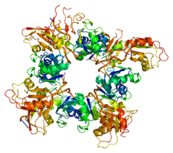
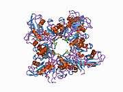
PDB gallery |
|---|
1cvj: X-RAY CRYSTAL STRUCTURE OF THE POLY(A)-BINDING PROTEIN IN COMPLEX WITH POLYADENYLATE RNA |