Nasal bone
| Nose bone | |
|---|---|
 Nasal bone visible at center, in dark green. | |
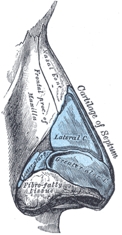 Cartilages of the nose. Side view. (Nasal bone visible at upper left.) | |
| Details | |
| Identifiers | |
| Latin | os nasale |
| MeSH | D009295 |
| TA | A02.1.10.001 |
| FMA | 52745 |
| Anatomical terms of bone | |
The nasal bones are two small oblong bones, varying in size and form in different individuals; they are placed side by side at the middle and upper part of the face and by their junction, form the bridge of the nose.
Each has two surfaces and four borders.
Structure
Surfaces
The outer surface is concavo-convex from above downward, convex from side to side; it is covered by the procerus and nasalis muscles, and perforated about its center by a foramen, for the transmission of a small vein.
The inner surface is concave from side to side, and is traversed from above downward, by a groove for the passage of a branch of the nasociliary nerve.
Articulations
The nasal articulates with four bones: two of the cranium, the frontal and ethmoid, and two of the face, the opposite nasal and the maxilla.
Other animals
In primitive bony fish and tetrapods, the nasal bones are the most anterior of a set of four paired bones forming the roof of the skull, being followed in sequence by the frontals, the parietals, and the postparietals. Their form in living species is highly variable, depending on the shape of the head, but they generally form the roof of the snout or beak, running from the nostrils to a position short of the orbits. In most animals, they are generally therefore proportionally larger than in humans or great apes, because of the shortened faces of the latter. Turtles, unusually, lack nasal bones, with the prefrontal bones of the orbit reaching all the way to the nostrils.[1]
Additional images
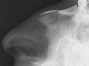 X-ray of nasal bone fracture
 Nasal bone
 Orbital bones
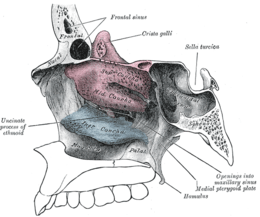 Lateral wall of nasal cavity, showing ethmoid bone in position.
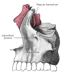 Articulation of nasal and lacrimal bones with maxilla.
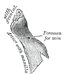 Right nasal bone. Outer surface.
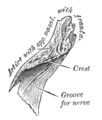 Right nasal bone. Inner surface.
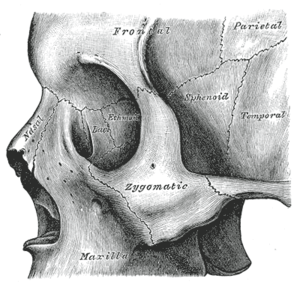 Close up of side view of the skull.
 Side view of the skull.
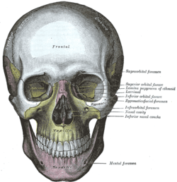 The skull from the front.
 Horizontal section of nasal and orbital cavities.
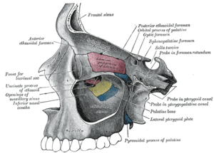 Medial wall of left orbit.
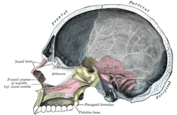 Sagittal section of skull.
 Roof, floor, and lateral wall of left nasal cavity.
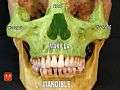 Nasal bone
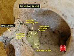 Lacrimal and nasal bones
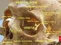 Nasal bone
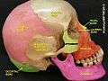 Nasal bone
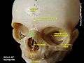 Nasal bone
See also
References
- This article incorporates text in the public domain from page 156 of the 20th edition of Gray's Anatomy (1918)
External links
| Wikimedia Commons has media related to Nasal bones. |
- Anatomy figure: 22:02-07 at Human Anatomy Online, SUNY Downstate Medical Center—"Anterior view of skull."
- Anatomy photo:29:st-0206 at the SUNY Downstate Medical Center—"Orbits and Eye: Bones"
- Anatomy figure: 33:01-03 at Human Anatomy Online, SUNY Downstate Medical Center—"The bones of the lateral nasal wall."
- "Anatomy diagram: 34256.000-1". Roche Lexicon - illustrated navigator. Elsevier. Archived from the original on 2014-01-01.