Mastitis in dairy cattle

Bovine mastitis is the persistent, inflammatory reaction of the udder tissue due to physical trauma or microorganisms infections. Mastitis, a potentially fatal mammary gland infection, is the most common disease in dairy cattle in the United States and worldwide. It is also the most costly disease to the dairy industry.[1] Milk from cows suffering from mastitis has an increased somatic cell count. Prevention and Control of mastitis requires consistency in sanitizing the cow ban facilities, proper milking procedure and segregation of infected animals. Treatment of the disease is carried out by penicillin injection in combination with sulphar drug.
Definition
Mastitis occurs when white blood cells (leukocytes) are released into the mammary gland, usually in response to bacteria invading the teat canal or occasionally by chemical, mechanical, or thermal trauma on the udder. Milk-secreting tissue and various ducts throughout the mammary gland are damaged due to toxins released by the bacteria resulting in reduced milk yield and quality.
Identification
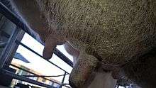
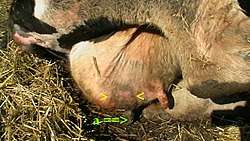
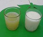
This disease can be identified by abnormalities in the udder such as swelling, heat, redness, hardness, or pain (if it is clinical). Other indications of mastitis may be abnormalities in milk such as a watery appearance, flakes, or clots. When infected with sub-clinical mastitis, a cow does not show any visible signs of infection or abnormalities in milk or on the udder.[1]
Mastitis-causing bacteria
Bacteria that are known to cause mastitis include:
- Pseudomonas aeruginosa
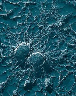
- Staphylococcus aureus
- Staphylococcus epidermidis
- Streptococcus agalactiae[2]
- Streptococcus uberis
- Brucella melitensis
- Corynebacterium bovis
- Mycoplasma spp. (including Mycoplasma bovis)
- Escherichia coli (E. coli)
- Klebsiella pneumoniae
- Klebsiella oxytoca
- Enterobacter aerogenes[3]
- Pasteurella spp.[4]
- Trueperella pyogenes[5] (previously Arcanobacterium pyogenes)
- Proteus spp.[6]
- Prototheca zopfii (achlorophyllic algae)
- Prototheca wickerhamii (achlorophyllic algae)[6]
These bacteria can be classified as environmental or contagious depending on mode and source of transmission.
Types of mastitis
Mastitis may be classified according two different criteria: either according to the clinical symptoms or depending on the mode of transmission.
- Clinical symptoms
- Clinical mastitis
- Sub-Clinical mastitis
- per acute mastitis
- acute mastitis
- sub acute mastitis
- chronic mastitis
- Mode of transmission
- Contagious mastitis
- environmental mastitis
Transmission
Mastitis is most often transmitted by repetitive contact with the milking machine, and through contaminated hands or materials.
Another route is via the oral-to-udder transmission among calves. Feeding calves on milk may introduce some mastitis causing bacteria strain in the oral cavity of the calf where it will stay dormant until it is transmitted elsewhere. Since grouped calves like to stimulate suckling, they will transmit the bacteria to the udder tissue of their fellow calves. The bacteria will lay dormant in the udder tissue as the calf grows until it begins to lactate. That is when the bacteria activates and causes mastitis. This calls for strict calf management practices to curb this route of transmission.
Effects on milk composition
Mastitis can cause a decline in potassium and an increase in lactoferrin. It also results in decreased casein, the major protein in milk. As most calcium in milk is associated with casein, the disruption of casein synthesis contributes to lowered calcium in milk. The milk protein continues to undergo further deterioration during processing and storage.[7] Milk from cows with mastitis also has a higher somatic cell count.[8] Generally speaking, the higher the somatic cell count, the lower the milk quality.
Management
Detection

Cattle affected by mastitis can be detected by examining the udder for inflammation and swelling, or by observing the consistency of the milk, which will often develop clots or change color when a cow is infected.[9]
Another method of detection is the California mastitis test, which is designed to measure the milk's somatic cell count as a means for detecting inflammation and infection of the udder.[10]
Treatment
Treatment is possible with long-acting antibiotics, but milk from such cows is not marketable until drug residues have left the cow's system. Antibiotics may be systemic (injected into the body), or they may be forced upwards into the teat through the teat canal (intramammary infusion). Cows being treated may be marked with tape to alert dairy workers, and their milk is syphoned off and discarded. To determine whether the levels of antibiotic residuals are within regulatory requirements, special tests exist. Vaccinations for mastitis are available, but as they only reduce the severity of the condition, and cannot prevent reoccurring infections, they should be used in conjunction with a mastitis prevention program.
Control
Practices such as good nutrition, proper milking hygiene, and the culling of chronically infected cows can help. Ensuring that cows have clean, dry bedding decreases the risk of infection and transmission. Dairy workers should wear rubber gloves while milking, and machines should be cleaned regularly to decrease the incidence of transmission.
Prevention
A good milking routine is vital. This usually consists of applying a pre-milking teat dip or spray, such as an iodine spray, and wiping teats dry prior to milking. The milking machine is then applied. After milking, the teats can be cleaned again to remove any growth medium for bacteria. A post milking product such as iodine-propylene glycol dip is used as a disinfectant and a barrier between the open teat and the bacteria in the air. Mastitis can occur after milking because the teat holes close after 15 minutes if the animal sits in a dirty place with feces and urine.
Industry costs
This disease costs the US dairy industry about 1.7 to 2 billion USD each year.[7]
References
- 1 2 Department of Animal Science. "Mastitis in Dairy Cows" (PDF). MacDonald Campus of McGill University. Archived from the original (PDF) on July 8, 2003. Retrieved 4 February 2010.
- ↑ "Teat Disinfection Facts". NMC. Retrieved 4 February 2010.
- ↑ "A Practical Look at Environmental Mastitis". .nmconline.org/. Retrieved 4 February 2010.
- ↑ "Mastitis Pathogen Notes: Pasteurella species". nmconline.org. Retrieved 4 February 2010.
- ↑ "Mastitis Pathogen Notes: Arcanobacterium pyogenes". nmconline.org. Retrieved 4 February 2010.
- 1 2 "Mastitis Pathogen Notes: Proteus species". nmconline.org. Retrieved 4 February 2010.
- 1 2 Jones, G. M.; Bailey, T. L. "Understanding the Basics of Mastitis". Virginia Cooperative Extension. Retrieved 4 February 2010.
- ↑ Kandasamy S, Green BB, Benjamin AL, Kerr DE. Between-cow variation in dermal fibroblast response to lipopolysaccharide reflected in resolution of inflammation during Escherichia coli mastitis. J Dairy Sci. 2011 Dec;94(12):5963-75. doi: 10.3168/jds.2011-4288. PubMed PMID 22118085
- ↑ Laven, Richard. "Mastitis Control and Management: Mastitis Part 4 - Detecting and Treating Clinical Mastitis". National Animal Disease Information Service. Retrieved 27 February 2015.
- ↑ "Detection of Mastitis". Department of Animal Sciences. University of Illinois at Urbana–Champaign. Retrieved 27 February 2015.
Further reading
- Harmon, R. J. 1994. Physiology of mastitis and factors affecting somatic cell counts. J. Dairy Sci. 77:2103-2112.
- Jones, G. M., R. E. Pearson, G. A. Clabaugh, and C. W. Heald. 1984. Relationships between somatic cell counts and milk production. J. Dairy Sci. 67:1823-1831.
- Myllys, V., and H. Rautala. 1995. Characterization of clinical mastitis in primiparous heifers. J. Dairy Sci. 78:538-545.
- National Mastitis Council. 1996. Current Concepts of Bovine Mastitis, 4th ed., Arlington, VA.
- Fox LK et al. Survey of intramammary infections in dairy heifers at breeding age and first parturition. J Dairy Sci. 78; 1619–1628, 1995.
- Hallberg JW et al. The visual appearance and somatic cell count of mammary secretions collected from primigravid heifers during gestation and early postpartum. J Dairy Sci. 78; 1629-1636.
- Hogan JS et al. Efficacy of an Escherichia coli J5 bacterin administered to primigravid heifers. J Dairy Sci. 82; 939-943, 1999.
- Nickerson SC. Mastitis and its control in heifers and dry cows. International Symposium on Bovine Mastitis. Indianapolis, IN, September, 1990. pp 82–91.
- Nickerson SC et al. Mastitis in dairy heifers: Initial studies on prevalence and control. J Dairy Sci. 78;1607–1618, 1995.
- Nickerson SC et al. Efficacy of s Staphylococcus aureus bacterin in dairy herifers. An update. Proceedings of the Nat Mastitis Council Meeting. 295-6, 1998.
- Sears PM and Wilson DJ. Heifer mastitis. Bov Practitioner 28; 56-58, 1994.