Hepatitis E virus
| Hepatitis E virus | |
|---|---|
 | |
| TEM micrograph of hepatitis E virions. | |
| Virus classification | |
| Group: | Group IV ((+)ssRNA) |
| Order: | Unassigned |
| Family: | Hepeviridae |
| Genus: | Orthohepevirus |
| Species: | Orthohepevirus A |
| Synonyms | |
| |
The hepatitis E virus, or HEV, is the causative agent of hepatitis E. Its taxonomic name is Orthohepevirus A.[1]
The global burden of infections from the two major genotypes (1 and 2) is estimated at 20 million per year, leading to 70,000 deaths and 3,000 stillbirths.[2]
The virus particle was first seen in 1983,[3] but was only molecularly cloned in 1989.[4]
Genome and proteome
HEV can be classified into eight different genotypes from different geographical regions: genotype 1 (Asia), genotype 2 (Africa and Mexico), genotype 3 (Europe and North America), genotype 4 (Asia); genotypes 5 and 6 have been detected in Asian wild boar and genotypes 7 and 8 in camels.[1][5]
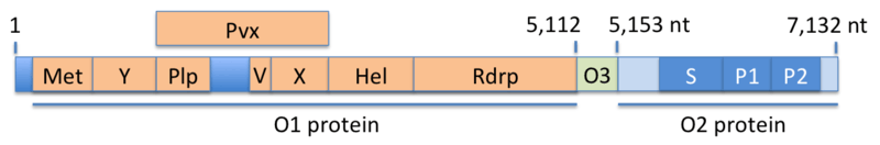
The viral genome a single strand of positive-sense RNA that is about 7200 bases in length. The three open-reading frames (ORF1, ORF2 and ORF3) encode for three proteins (O1, O2, O3), two of which are polyproteins, that is, they are cleaved into fragments which carry out the actual functions of the virus (see figure). The O1 protein consists of seven such fragments, namely Met (methyltransferase), Y (Y-domain), Plp (papain-like protease), V (proline-rich variable region), X (X-domain, macro-domain), Hel (helicase), and Rdrp (RNA-dependent RNA polymerase). The Pvx domain is a fusion protein consisting of the Plp, V, and X domains. The O3 protein is encoded by a single open-reading frame (ORF3). The O2 protein encodes the capsid, which is composed of three domains, namely the shell domain (S) and two protruding domains (P1, P2).[6] Numbers in the figure indicate positions in the RNA sequence.
Interactome
The protein-protein interactome among HEV proteins has been mapped by Osterman et al. (2015), who found 25 interactions among the 10 proteins studied. Almost all (24) of these interactions were considered as of "high quality".[7]
Structure
The viral particles are 27 to 34 nanometers in diameter and are not enveloped.[1][3]
Taxonomy
It was previously classified in the Caliciviridae family. However, its genome more closely resembles the rubella virus. It is now classified as a member of the genus Orthohepevirus in the Hepeviridae family.[1]
See also
References
- 1 2 3 4 "ICTV Online (10th) Report".
- ↑ Rein, D. B., Stevens, G. A., Theaker, J., Wittenborn, J. S. & Wiersma, S. T. (2012) The global burden of hepatitis E virus genotypes 1 and 2 in 2005. Hepatology 55, 988–97
- 1 2 Balayan MS, Andjaparidze AG, Savinskaya SS, et al. (1983). "Evidence for a virus in non-A, non-B hepatitis transmitted via the fecal-oral route". Intervirology. 20 (1): 23–31. doi:10.1159/000149370. PMID 6409836.
- ↑ Reyes GR, Purdy MA, Kim JP, et al. (1990). "Isolation of a cDNA from the virus responsible for enterically transmitted non-A, non-B hepatitis". Science. 247 (4948): 1335–9. doi:10.1126/science.2107574. PMID 2107574.
- ↑ Schlauder, G. G. & Mushahwar, I. K. (2001) Genetic heterogeneity of hepatitis E virus. J Med Virol 65, 282–92
- ↑ Ahmad I, Holla RP, Jameel S (2011). "Molecular virology of hepatitis E virus". Virus Res. 161 (1): 47–58. doi:10.1016/j.virusres.2011.02.011. PMC 3130092. PMID 21345356.
- ↑ Osterman A, Stellberger T, Gebhardt A, Kurz M, Friedel CC, Uetz P, Nitschko H, Baiker A, Vizoso-Pinto MG (2015). "The Hepatitis E virus intraviral interactome". Sci Rep. 5: 13872. doi:10.1038/srep13872. PMC 4604457. PMID 26463011.
External links
- ICTV Online (10th) Report Hepeviridae
- Hepatitis+E+virus at the US National Library of Medicine Medical Subject Headings (MeSH)
- Virus Pathogen Database and Analysis Resource (ViPR): Hepeviridae
- "Orthohepevirus", Catalan Wikipedia (written by Veterinarian Students of the University Autonomy of Barcelona, Spain).