Interventional radiology
| Interventional radiology | |
|---|---|
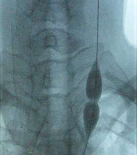 Balloon dilatation of the stenosed internal jugular vein (photo from an X-ray angiograph monitor). While pressure in the balloon is relatively low, stenosis prevents the balloon from inflating in the middle. Further increase in pressure will dilate the narrowing and restore the full blood flow. | |
| MeSH | D015642 |
Interventional radiology (IR), sometimes known as vascular and interventional radiology (VIR), is a medical specialty which provides minimally invasive image-guided diagnosis and treatment of disease. Although the range of procedures performed by interventional radiologists is broad, the unifying concept behind these procedures is the application of image guidance and minimally invasive techniques in order to minimize risk to the patient.
Training
| Occupation | |
|---|---|
| Names | Doctor, Medical Specialist |
Occupation type | Specialty |
Activity sectors | Medicine |
| Description | |
Education required |
|
Fields of employment | Hospitals, Clinics |
Related jobs | Radiologist |
United States
Training for interventional radiology occurs in the residency portion of medical education, and has gone through developments.
In 2000, the Society of Interventional Radiology (SIR) created a program named "Clinical Pathway in IR", which modified the "Holman Pathway" that was already accepted by the American Board of Radiology to including training in IR; this was accepted by ABR but was not widely adopted. In 2005 SIR proposed and ABR accepted another pathway called "DIRECT (Diagnostic and Interventional Radiology Enhanced Clinical Training) Pathway" to help trainees coming from other specialities learn IR; this too was not widely adopted. In 2006 SIR proposed a pathway resulting in certification in IR as a speciality; this was eventually accepted by the ABR in 2007 and was presented to the American Board of Medical Specialities (ABMS) in 2009, which rejected it because it did not include enough diagnostic radiology (DR) training. The proposal was reworked, at the same time that overall DR training was being revamped, and a new proposal that would lead to a dual DR/IR specialization was presented to the ABMS and was accepted in 2012 and eventually was implemented in 2014.[1][2][3] By 2016 the field had determined that the old IR fellowships would be terminated by 2020.[3]
A handful of programs have offered interventional radiology fellowships that focus on training in the treatment of children.[4]
Europe
In Europe the field followed its own pathway; for example in Germany the parallel interventional society began to break free of the DR society in 2008.[5] In the UK, interventional radiology was approved as a sub-specialty of clinical radiology in 2010.[6][7] While many countries have an interventional radiology society, there is also the European-wide Cardiovascular and Interventional Radiological Society of Europe, whose aim is to support teaching, science, research and clinical practice in the field by hosting meetings, educational workshops and promoting patient safety initiatives. Furthermore, the Society provides an examination, the European Board of Interventional Radiology (EBIR), which is a highly valuable qualification in interventional radiology based on the European Curriculum and Syllabus for IR.
Procedures
Interventional radiologists commonly perform both diagnostic and therapeutic procedures, although diagnostic angiography is becoming less common as the quality and reliability of CT and MRI angiography has allowed for alternative forms of non invasive evaluation.
Diagnostic
- Angiography: Imaging the blood vessels to look for abnormalities with the use of various contrast media, including iodinated contrast, gadolinium based agents, and CO2 gas.[8]
- Cholangiography: Imaging the bile ducts within the liver to look for areas of blockage.
- Biopsy: Taking of a tissue sample from the area of interest for pathological examination from a percutaneous or transvenous approach.[9]
Therapeutic
Vascular
- Balloon angioplasty/stent: Opening of narrow or blocked blood vessels using a balloon, with or without placement of metallic stents to aid in keep vessel patent.[10]
- Endovascular aneurysm repair: Placement of endovascular stent-graft across an aneurysm, in order to prevent expansion or progression of the defective vessel.[11]
- Embolization: Placement of a metallic coil or embolic substance (gel-foam, poly-vinyl alcohol) to block blood through to a blood vessel, either to stop bleeding or decrease blood flow to a target organ or tissue.[12]
- Uterine artery embolization (UAE) or uterine fibroid embolization (UFE)
- Prostate artery embolization (PAE)
- Thrombolysis: Catheter-directed technique for dissolving blood clots, such as pulmonary embolism, deep venous thrombosis) with either pharmaceutical (TPA) or mechanical means.
- IVC filters: Metallic filters placed in the vena cava to prevent propagation of deep venous thrombus.
- Dialysis related interventions: Placement of tunneled hemodialysis catheters, peritoneal dialysis catheters, and revision/thrombolysis of poorly functioning surgically placed AV fistulas and grafts.[13]
- TIPS: Placement of a Transjugular Intrahepatic Porto-systemic Shunt (TIPS) for select indications in patients with critical end-stage liver disease and portal hypertension.[14]
- Endovenous laser treatment of varicose veins: Placement of thin laser fiber in varicose veins for non-surgical treatment of venous insufficiency.
- Placement of catheters in the biliary system to bypass biliary obstructions and decompress the biliary system.
- Placement of permanent indwelling biliary stents.
- Cholecystostomy: Placement of a tube into the gallbladder to remove infected bile in patients with cholecystitis, an inflammation of the gallbladder, who are too frail or too sick to undergo surgery.
Catheter placement
- Central venous catheter placement: Vascular access and management of intravenous devices (IVs), including both tunneled and non-tunneled catheters (e.g. PIC, Hickman, port catheters, hemodialysis catheters, translumbar and transhepatic venous lines).
- Drainage catheter placement: Placement of tubes to drain pathologic fluid collections (e.g., abscess, pleural effusion). This may be achieved by percutaneous, trans-rectal, or trans-vaginal approach. Exchange or repositioning of indwelling catheters is achieved over a guidewire under image guidance.
- Radiologically inserted gastrostomy or jejunostomy : Placement of a feeding tube percutaneously into the stomach and/or jejunum.[16]
- Chemoembolization: combined injection of chemotherapy and embolic agents into the arterial blood supply of a tumor, with the goal of both local administration of chemotherapy, slowing "washout" of the chemotherapy drug, and also decreasing tumor arterial supply.
- Radioembolization: combined injection of radioactive glass or plastic beads and embolic agents into the arterial blood supply of a tumor, with the goal of both local administration of radiotherapy, slowing "washout" of the radioactive substance, and also decreasing tumor arterial supply.
- Radiofrequency ablation (RF/RFA): local treatment which uses a special catheter to destroy tissue by using heat generated by medium frequency alternating currents.
- Cryoablation: local treatment which uses a special catheter to destroy tissue by using cold temperature generated by rapid expansion of compressed argon gas. This technique is mostly used for the treatment of small renal cancers and for the palliation of painful bone lesions.[19]
- Microwave ablation: local treatment which uses a special catheter to destroy tissue by using heat generated by microwaves.
Genitourinary[20]
- Percutaneous nephrostomy or nephroureteral stent placement: Placement of a catheter through the skin, directly into the kidney in order to drain from the collecting system. This is typically done to treat a downstream obstruction of urine.
- Ureteral stent exchange: indwelling double-J type ureteral stents, typically placed by urologist using cystoscopy, may be exchanged in retrograde fashion through the female urethra. The IR uses a thin wire snare under fluoroscopy to capture the distal portion of the stent. After partially extracting the distalmost stent, exchange for a new stent can be accomplished over a guidewire.
Pain management
- Vertebroplasty: Percutaneous injection of biocompatible bone cement inside a fractured spinal vertebrae in order to restore vertebral body height and relieve pain.[21]
References
- ↑ Kaufman, John A. (November 2014). "The Interventional Radiology/Diagnostic Radiology Certificate and Interventional Radiology Residency". Radiology. 273 (2): 318–321. doi:10.1148/radiol.14141263. PMID 25340266.
- ↑ Siragusa, DA; Cardella, JF; Hieb, RA; Kaufman, JA; Kim, HS; Nikolic, B; Misra, S; Resnick, SA; Saad, WE; Vatakencherry, G; Wallace, MJ; Society of Interventional, Radiology. (November 2013). "Requirements for training in interventional radiology". Journal of vascular and interventional radiology : JVIR. 24 (11): 1609–12. doi:10.1016/j.jvir.2013.08.002. PMC 4485607. PMID 24160820.
- 1 2 Di Marco, L; Anderson, MB (February 2016). "The new Interventional Radiology/Diagnostic Radiology dual certificate: "higher standards, better education"". Insights into imaging. 7 (1): 163–5. doi:10.1007/s13244-015-0450-9. PMC 4729716. PMID 26746975.
- ↑ The Society for Pediatric Radiology Archived 2012-01-04 at the Wayback Machine.
- ↑ Mahnken, AH; Bücker, A; Hohl, C; Berlis, A (April 2017). "White Paper: Curriculum in Interventional Radiology". RoFo : Fortschritte auf dem Gebiete der Rontgenstrahlen und der Nuklearmedizin. 189 (4): 309–311. doi:10.1055/s-0043-104773. PMID 28335057.
- ↑ Kassamali, Rahil H.; Hoey, Edward T.D. (December 2014). "Radiology training in United Kingdom: current status". Quantitative Imaging in Medicine and Surgery. 4 (6): 447–448. doi:10.3978/j.issn.2223-4292.2014.10.10. PMC 4256234. PMID 25525574.
- ↑ "Guidance on Training in Interventional Radiology" (PDF). Royal College of Radiologists. Retrieved 26 September 2017.
- ↑ Uberoi, Raman (2009). "4 Imaging". Interventional radiology. Oxford New York: Oxford University Press. pp. 49–77. ISBN 978-0-19-157556-3.
- ↑ Uberoi, Raman (2009). "19 Biopsy and drainage". Interventional radiology. Oxford New York: Oxford University Press. pp. 387–402. ISBN 978-0-19-157556-3.
- ↑ Uberoi, Raman (2009). "7 Angioplasty and stenting". Interventional radiology. Oxford New York: Oxford University Press. pp. 123–147. ISBN 978-0-19-157556-3.
- ↑ Uberoi, Raman (2009). "9 Stentgrafting". Interventional radiology. Oxford New York: Oxford University Press. pp. 171–186. ISBN 978-0-19-157556-3.
- ↑ Uberoi, Raman (2009). "17 Embolization techniques". Interventional radiology. Oxford New York: Oxford University Press. pp. 341–360. ISBN 978-0-19-157556-3.
- ↑ Uberoi, Raman (2009). "12 Haemodialysis fistula". Interventional radiology. Oxford New York: Oxford University Press. pp. 253–268. ISBN 978-0-19-157556-3.
- ↑ Keller, Frederick S.; Farsad, Khashayar; Rösch, Josef (2016). "The Transjugular Intrahepatic Portosystemic Shunt: Technique and Instruments". Techniques in vascular and interventional radiology. Elsevier BV. 19 (1): 2–9. doi:10.1053/j.tvir.2016.01.001. ISSN 1089-2516. PMID 26997084.
- ↑ Uberoi, Raman (2009). "13 Hepatobiliary intervention". Interventional radiology. Oxford New York: Oxford University Press. pp. 269–282. ISBN 978-0-19-157556-3.
- ↑ Uberoi, Raman (2009). "14 Gastro-intestinal intervention". Interventional radiology. Oxford New York: Oxford University Press. pp. 290–295. ISBN 978-0-19-157556-3.
- ↑ Uberoi, Raman (2009). "18 Tumour ablation". Interventional radiology. Oxford New York: Oxford University Press. pp. 361–386. ISBN 978-0-19-157556-3.
- ↑ Wah, T.M. (2017). "Image-guided ablation of renal cell carcinoma". Clinical radiology. Elsevier BV. 72 (8): 636–644. doi:10.1016/j.crad.2017.03.007. ISSN 0009-9260. PMID 28527529.
- ↑ Percutaneous Tumor Ablation. Thieme Verlag. 2011. doi:10.1055/b-0034-81500. ISBN 9781604063066.
- ↑ Uberoi, Raman (2009). "11 Interventional uro-radiology". Interventional radiology. Oxford New York: Oxford University Press. pp. 221–225. ISBN 978-0-19-157556-3.
- ↑ Uberoi, Raman (2009). "22 Musculoskeletal intervention". Interventional radiology. Oxford New York: Oxford University Press. p. 445. ISBN 978-0-19-157556-3.
Further reading
- Historic Highlights of Interventional Radiology, by Josef Rösch of Dotter Interventional Radiology.
- Abrams’ Angiography: Vascular and Interventional Radiology. Herbert L. Abrams (Editor), Stanley Baum (Editor) and Michael J. Pentecost (Editor). Little Brown and Co., 2005. ISBN 0781740894
- Advanced Radiographic and Angiographic Procedures: With an Introduction to Specialized Imaging. Patrick A. Apfel, Marianne Rita Tortorici. F A Davis Co., 2010. ISBN 0803612559
- Handbook of Interventional Radiologic Procedures Krishna Kandarpa (Editor) and John E. Aruny (Editor). Lippincott Williams and Wilkins Publishers, 2010. ISBN 0781768160
- Rösch Josef; Keller Frederick S.; Kaufman John A. (2003). "The Birth, Early Years, and Future of Interventional Radiology" (PDF). J. Vasc. Interv. Radiol. 14: 841–853. doi:10.1097/01.rvi.0000083840.97061.5b. PMID 12847192.
- The Catheter Introducers by Leslie A. Geddes and LaNelle E. Geddes of Cook Group Incorporated, Mobium Press, Chicago. 1993. ISBN 0916371131
- The Ship in the Balloon: The Story of Boston Scientific and the Development of Less-Invasive Medicine by Jeffrey L. Rodengen. Write Stuff Enterprises, Inc., Fr Lauderdale. 2001. ISBN 0945903502
External links
- Society of Interventional Radiology
- Cardiovascular and Interventional Radiological Society of Europe
- Society of Pediatric Interventional Radiology
- Interventional radiology procedures