Fluctuation X-ray scattering

Fluctuation X-ray scattering (FXS)[1][2] is an X-ray scattering technique similar to small-angle X-ray scattering (SAXS), but is performed using X-ray exposures below sample rotational diffusion times. This technique, ideally performed with an ultra-bright X-ray light source, such as a free electron laser, results in data containing significantly more information as compared to traditional scattering methods.[3]
FXS can be used for the determination of (large) macromolecular structures,[4] but has also found applications in the characterization of metallic nanostructures,[5] magnetic domains[6] and colloids.[7]
The most general setup of FXS is a situation in which fast diffraction snapshots of models are taken which over a long time period undergo a full 3D rotation. A particularly interesting subclass of FXS is the 2D case where the sample can be viewed as a 2-dimensional system with particles exhibiting random in-plane rotations. In this case, an analytical solution exists relation the FXS data to the structure.[8] In absence of symmetry constraints, no analytical data-to-structure relation for the 3D case is available, although various iterative procedures have been developed.
Overview
An FXS experiment consists of collecting a large number of X-ray snapshots of samples in a different random configuration. By computing angular intensity correlations for each image and averaging these over all snapshots, the average 2-point correlation function can be subjected to a finite Legendre transform, resulting in a collection of so-called Bl(q,q') curves, where l is the Legendre polynomial order and q / q' the momentum transfer or inverse resolution of the data.
Mathematical background
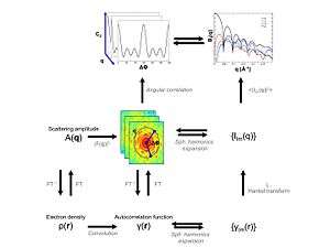
Given a particle : , the associated three-dimensional complex structure factor : is obtained via a Fourier transform


![{\displaystyle A(\mathbf {q} )=\int _{V}\rho (\mathbf {r} )\exp[i\mathbf {qr} ]d\mathbf {r} }](../I/m/ca830e3f8b40b966e7e046af1b1fe439f7245c3b.svg)
The intensity function corresponding to the complex structure factor is equal to

where : denotes complex conjugation. Expressing : as a spherical harmonics series, one obtains


The average angular intensity correlation as obtained from many diffraction images : is then


It can be shown that:
![{\displaystyle C_{2}(q,q',\Delta \phi _{q})=\sum _{l}B_{l}(q,q')P_{l}(\cos(\theta _{q})\cos(\theta _{q'})+\sin(\theta _{q})\sin(\theta _{q'})\cos[\Delta \phi _{q}])}](../I/m/8c5c8dd9060880ede71a80ae0e90ffea3d4f38ed.svg)
where

with : equal to the X-ray wavelength used, and


- is a Legendre Polynome. The set of : curves can be obtained via a finite Legendre transform from the observed autocorrelation : and are thus directly related to the structure : via the above expressions.


Additional relations can be obtained by obtaining the real space autocorrelation : of the density:


A subsequent expansion of : in a spherical harmonics series, results in radial expansion coefficients that are related to the intensity function via a Hankel transform

A concise overview of these relations has been published elsewhere[1][3]
Basic relations
A generalized Guinier law describing the low resolution behavior of the data can be derived from the above expressions:

Values of : and : can be obtained from a least squares analyses of the low resolution data.[3]


The falloff of the data at higher resolution is governed by Porod laws. It can be shown [3] that the Porod laws derived for SAXS/WAXS data hold here as well, ultimately resulting in:

for particles with well-defined interfaces.
Structure determination from FXS data
Currently, there are three routes to determine molecular structure from its corresponding FXS data.
Algebraic phasing
By assuming a specific symmetric configuration of the final model, relations between expansion coefficients describing the scattering pattern of the underlying species can be exploited to determine a diffraction pattern consistent with the measure correlation data. This approach has been shown to be feasible for icosahedral[9] and helical models.[10]
Reverse Monte Carlo
By representing the to-be-determined structure as an assembly of independent scattering voxels, structure determination from FXS data is transformed into a global optimisation problem and can be solved using simulated annealing.[3]
Multi-tiered iterative phasing
The multi-tiered iterative phasing algorithm (M-TIP) overcomes convergence issues associated with the reverse Monte Carlo procedure and eliminates the need to use or derive specific symmetry constraints as needed by the Algebraic method. The M-TIP algorithm utilizes non-trivial projections that modifies a set of trial structure factors : such that corresponding : match observed values. The real-space image : , as obtained by a Fourier Transform of : is subsequently modified to enforce symmetry, positivity and compactness. The M-TIP procedure can start from a random point and has good convergence properties.[11]
References
- 1 2 Kam, Zvi (1977). "Determination of Macromolecular Structure in Solution by Spatial Correlation of Scattering Fluctuations". Macromolecules. 10 (5): 927–934. Bibcode:1977MaMol..10..927K. doi:10.1021/ma60059a009.
- ↑ Kam, Z.; M. H. Koch, and J. Bordas (1981). "Fluctuation x-ray scattering from biological particles in frozen solution by using synchrotron radiation". Proceedings of the National Academy of Sciences of the United States of America. 78 (6): 3559–3562. Bibcode:1981PNAS...78.3559K. doi:10.1073/pnas.78.6.3559. PMC 319609.
- 1 2 3 4 5 6 Malmerberg, Erik; Cheryl A. Kerfeld and Petrus H. Zwart (2015). "Operational properties of fluctuation X-ray scattering data". IUCrJ. 2 (3): 309–316. doi:10.1107/S2052252515002535. PMC 4420540. PMID 25995839.
- ↑ Liu, Haiguang; Poon, Billy K.; Saldin, Dilano K.; Spence, John C. H.; Zwart, Peter H. (2013). "Three-dimensional single-particle imaging using angular correlations from X-ray laser data". Acta Crystallographica Section A. 69 (4): 365–373. doi:10.1107/S0108767313006016. ISSN 0108-7673.
- ↑ Chen, Gang; Modestino, Miguel A.; Poon, Billy K.; Schirotzek, André; Marchesini, Stefano; Segalman, Rachel A.; Hexemer, Alexander; Zwart, Peter H. (2012). "Structure determination of Pt-coated Au dumbbellsviafluctuation X-ray scattering". Journal of Synchrotron Radiation. 19 (5): 695–700. doi:10.1107/S0909049512023801. ISSN 0909-0495.
- ↑ Su, Run; Seu, Keoki A.; Parks, Daniel; Kan, Jimmy J.; Fullerton, Eric E.; Roy, Sujoy; Kevan, Stephen D. (2011). "Emergent Rotational Symmetries in Disordered Magnetic Domain Patterns". Physical Review Letters. 107 (25): 257204. Bibcode:2011PhRvL.107y7204S. doi:10.1103/PhysRevLett.107.257204. ISSN 0031-9007. PMID 22243108.
- ↑ Wochner, Peter; Gutt, Christian; Autenrieth, Tina; Demmer, Thomas; Bugaev, Volodymyr; Ortiz, Alejandro Díaz; Duri, Agnès; Zontone, Federico; Grübel, Gerhard; Dosch, Helmut (2009). "X-ray cross correlation analysis uncovers hidden local symmetries in disordered matter". Proceedings of the National Academy of Sciences. 106 (28): 11511–11514. Bibcode:2009PNAS..10611511W. doi:10.1073/pnas.0905337106. ISSN 0027-8424. PMC 2703671. PMID 20716512.
- ↑ Kurta, R. P.; Altarelli, M.; Weckert, E.; Vartanyants, I. A. (2012). "X-ray cross-correlation analysis applied to disordered two-dimensional systems". Physical Review B. 85 (18). arXiv:1202.6253. Bibcode:2012PhRvB..85r4204K. doi:10.1103/PhysRevB.85.184204. ISSN 1098-0121.
- ↑ Saldin, D. K.; H.-C. Poon, P. Schwander, M. Uddin, and M. Schmidt (2011). "Reconstructing an icosahedral virus from single-particle diffraction experiments". Optics Express. 19 (18): 17318–17335. arXiv:1107.5212. Bibcode:2011OExpr..1917318S. doi:10.1364/OE.19.017318.
- ↑ Poon, H.-C.; P. Schwander, M. Uddin, & D. K. Saldin (2011). "Fiber Diffraction without Fibers" (PDF). Physical Review Letters. 19 (18): 17318–17335. Bibcode:2013PhRvL.110z5505P. doi:10.1103/PhysRevLett.110.265505.
- ↑ Donatelli, Jeffrey J.; Peter H. Zwart, and James A. Sethian (2015). "Iterative phasing for fluctuation X-ray scattering" (PDF). Proceedings of the National Academy of Sciences of the United States of America. 112 (33): 10286–10291. Bibcode:2015PNAS..11210286D. doi:10.1073/pnas.1513738112. PMC 4547282. PMID 26240348. early edition online ahead of publication