Diverticulum
| Diverticulum | |
|---|---|
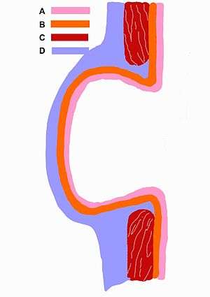 | |
| Schematic drawing of a false diverticulum. A - mucosa; B - submucosa; C - muscularis; D - serosa and subserosa | |
| Classification and external resources | |
| MeSH | D004240 |
A diverticulum (plural: diverticula) is the medical or biological term for an outpouching of a hollow (or a fluid-filled) structure in the body. Depending upon which layers of the structure are involved, they are described as being either true or false.
In medicine, the term usually implies the structure is not normally present. However, in the embryonic stage, some normal structures begin development as a diverticulum arising from another structure.
Anatomical
Guttural pouch: A large (300-500 ml), paired, air-filled ventral diverticulum of the auditory tube found in horses and other Perissodactyla.
Classification
Diverticula are described as being true or false depending upon the layers involved:
- True diverticula involve all layers of the structure, including muscularis propria and adventitia, such as Meckel's diverticulum.
- False diverticula (also known as "pseudodiverticula") do not involve muscular layers or adventitia. False diverticula, in the GI tract for instance, involve only the submucosa and mucosa.
Human pathology
- Bladder diverticulum: Balloon-like growths on the bladder commonly associated with a chronic outflow obstruction, such as benign prostatic hyperplasia in older males. Usually found in pairs on opposite sides of the bladder, bladder diverticula are often surgically removed to prevent infection, rupture, or even cancer.
- Cardiac diverticulum: A very rare congenital malformation of the heart that is usually benign [1]
- Colonic diverticula: These can become infected (see diverticulitis) and can perforate, requiring surgery
- Diverticulum of Kommerell: unusual nomenclature, in that focal dilatations of a blood vessel are properly referred to as aneurysms
- Duodenal and jejunal diverticul(um|a): congenital lesions, may be a source of bacterial overgrowth, may perforate and may result in abscesses
- Epiphrenic diverticulum: due to dysfunction of the lower esophageal sphincter, as in achalasia
Diverticula may occur in one of the three areas of the esophagus - the pharyngoesophageal, the midesophageal area or the epiphrenic area of esophagus. Zenker's diverticulum is found three times more frequently in men than in women. It occurs posteriorly through the cricopharyngeal muscle in the midline of the neck. Usually seen in people older than 60 years of age.
- Gastric diverticula - "Although usually asymptomatic, they may cause vague epigastric pain. These lesions may be confused radiologically for gastric ulcers or cancers. Endoscopically, they may be confused for paraesophageal hernias."[2]
- Killian-Jamieson diverticulum
- Meckel's diverticulum: a persistent portion of the omphalomesenteric duct present in 2% of the population
- Rokitansky-Aschoff sinuses: in the gallbladder due to chronic cholecystitis[3]
- Traction esophageal diverticulum: due to scarring from mediastinal or pulmonary tuberculosis
- Urethral diverticulum: congenital in males, post-infectious in females
- Zenker's diverticulum: a diverticulum of the mucosa of the pharynx affecting adults
Most of these pathological types of diverticulum are capable of harboring an enterolith. If the enterolith stays in place, it may cause no problems, but a large enterolith expelled from a diverticulum into the lumen can cause obstruction.
 Diverticulum of urinary bladder of a 59-year-old man, transverse plane
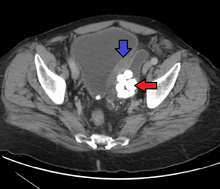 Bladder diverticula containing stones. Also note that the bladder wall is thickened due to possible transitional cell carcinoma.
 Large bowel (sigmoid colon) showing multiple diverticula. Note how the diverticula appear on either side of the longitudinal muscle bundle (taenium).
Embryological
- The kidneys, originally diverticula in the development of the urinary and reproductive organs
- The lungs, originally diverticula forming off of the ventral foregut.
- The thymus appears in the form of two flask-shape diverticula, which arise from the third branchial pouch (pharyngeal pouch) of the endoderm.
See also
Footnotes
- ↑ Vazquez-Jimenez, Dr. Jaime (2003). "Cardiac diverticulum" (PDF). Orphanet Encyclopedia. Retrieved 2008-01-14.
- ↑ Velanovich, V. (1994). "Gastric diverticulum". Surgical Endoscopy. 8 (11): 1338–1339. doi:10.1007/BF00188296. PMID 7831610.
- ↑ Stunell, H; Buckley, O; Geoghegan, T; O’Brien, J; Ward, E; Torreggiani, W (2008). "Imaging of adenomyomatosis of the gall bladder". Journal of Medical Imaging and Radiation Oncology. 52 (2): 109–117. doi:10.1111/j.1440-1673.2008.01926.x. ISSN 1754-9477.
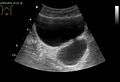
- 1 2 "UOTW #56 - Ultrasound of the Week". Ultrasound of the Week. 21 August 2015.