DNA beta-glucosyltransferase
| DNA beta-glucosyltransferase | |||||||||
|---|---|---|---|---|---|---|---|---|---|
| Identifiers | |||||||||
| EC number | 2.4.1.27 | ||||||||
| CAS number | 9030-14-2 | ||||||||
| Databases | |||||||||
| IntEnz | IntEnz view | ||||||||
| BRENDA | BRENDA entry | ||||||||
| ExPASy | NiceZyme view | ||||||||
| KEGG | KEGG entry | ||||||||
| MetaCyc | metabolic pathway | ||||||||
| PRIAM | profile | ||||||||
| PDB structures | RCSB PDB PDBe PDBsum | ||||||||
| |||||||||
In enzymology, a DNA beta-glucosyltransferase (EC 2.4.1.27) is an enzyme that catalyzes the chemical reaction in which a beta-D-glucosyl residue is transferred from UDP-glucose to an hydroxymethylcytosine residue in DNA. It is analogous to the enzyme DNA alpha-glucosyltransferase.
This enzyme belongs to the family of glycosyltransferases, specifically the hexosyltransferases. The systematic name of this enzyme class is UDP-glucose:DNA beta-D-glucosyltransferase. Other names in common use include T4-HMC-beta-glucosyl transferase, T4-beta-glucosyl transferase, T4 phage beta-glucosyltransferase, UDP glucose-DNA beta-glucosyltransferase, and uridine diphosphoglucose-deoxyribonucleate beta-glucosyltransferase.
Structural studies
As of late 2007, 20 structures have been solved for this class of enzymes, with PDB accession codes 1BGT, 1BGU, 1C3J, 1IXY, 1J39, 1JEJ, 1JG6, 1JG7, 1JIU, 1JIV, 1JIX, 1M5R, 1NVK, 1NZD, 1NZF, 1QKJ, 1SXP, 1SXQ, 2BGT, and 2BGU.
Bacteriophage T4 beta-glucosyltransferase
| T4-Gluco-transf | |||||||||
|---|---|---|---|---|---|---|---|---|---|
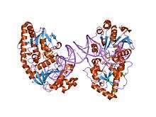 ternary complex of t4 phage bgt with udp and a 13 mer dna duplex | |||||||||
| Identifiers | |||||||||
| Symbol | T4-Gluco-transf | ||||||||
| Pfam | PF09198 | ||||||||
| InterPro | IPR015281 | ||||||||
| SCOP | 1jix | ||||||||
| SUPERFAMILY | 1jix | ||||||||
| |||||||||
In molecular biology,Bacteriophage T4 beta-glucosyltransferase refers to a protein domain found in a virus of Escherichia coli named bacteriophage T4. Members of this family are enzymes encoded by bacteriophage T4, which modify DNA by transferring glucose from uridine diphosphoglucose to 5-hydroxymethyl cytosine bases of phage T4 DNA.[1]
Function
Beta-glucosyltransferase is an enzyme, or more specifically an inverting glycosyltransferase(GT). In other words, it transfers glucose from uridine diphospho-glucose (UDPglucose)to an acceptor, modified DNA through beta-Glycosidic bond. The role of the enzyme is to protect the infecting viral DNA from the bacteria's restriction enzymes. Glucosylation prevents the virus DNA from being cut up. Furthermore, glucosylation may aid gene expression of the bacteriophage by influencing transcription.[2][3][4]
Structure
This structure has both alpha helices and beta strands.[2]
References
- ↑ Moréra S, Larivière L, Kurzeck J, Aschke-Sonnenborn U, Freemont PS, Janin J, Rüger W (August 2001). "High resolution crystal structures of T4 phage beta-glucosyltransferase: induced fit and effect of substrate and metal binding". J. Mol. Biol. 311 (3): 569–77. doi:10.1006/jmbi.2001.4905. PMID 11493010.
- 1 2 Larivière L, Gueguen-Chaignon V, Moréra S (2003). "Crystal structures of the T4 phage beta-glucosyltransferase and the D100A mutant in complex with UDP-glucose: glucose binding and identification of the catalytic base for a direct displacement mechanism". J Mol Biol. 330 (5): 1077–86. doi:10.1016/s0022-2836(03)00635-1. PMID 12860129.
- ↑ Moréra S, Imberty A, Aschke-Sonnenborn U, Rüger W, Freemont PS (1999). "T4 phage beta-glucosyltransferase: substrate binding and proposed catalytic mechanism". J Mol Biol. 292 (3): 717–30. doi:10.1006/jmbi.1999.3094. PMID 10497034.
- ↑ Moréra S, Larivière L, Kurzeck J, Aschke-Sonnenborn U, Freemont PS, Janin J, et al. (2001). "High resolution crystal structures of T4 phage beta-glucosyltransferase: induced fit and effect of substrate and metal binding". J Mol Biol. 311 (3): 569–77. doi:10.1006/jmbi.2001.4905. PMID 11493010.
- Kornberg SR, Zimmerman SB & Kornberg A (1961). "Glucosylation of deoxyribonucleic acid by enzymes from bacteriophage-infected Escherichia coli". J. Biol. Chem. 236: 1487&ndash, 1493.