Claustrum
| Claustrum | |
|---|---|
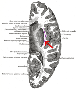
 Coronal section of human cerebrum. The claustrum is indicated by the arrow. | |
 Transverse section of human cerebrum. The claustrum is indicated by the arrow. | |
| Identifiers | |
| NeuroNames | 252 |
| NeuroLex ID | birnlex_1522 |
| TA | A14.1.09.421 |
| FMA | 76440 |
| Anatomical terms of neuroanatomy | |
The claustrum is a thin, irregular sheet of neurons that is attached to the underside of the neocortex in the center of the brain. It is suspected to be present in the brains of all mammals.
The claustrum in the human brain is a fraction of a millimetre to a few millimetres thick and is a vertical curved sheet of subcortical gray matter oriented sagittally between the white matter tracts of the external capsule and extreme capsule. The claustrum is lateral to the putamen and medial to the insular cortex and is considered by some sources to be part of the basal ganglia. There are lateral and medial tracts connecting the claustrum to many parts of the cortex and perhaps to the hippocampus, the amygdala, and the caudate nucleus (connections with subcortical centers are a matter of debate).
The claustrum has a uniformity in its types of cells, indicating a uniform type of processing by all claustral neurons. Though organized into modality specific regions, the claustrum contains a great deal of longitudinal connections between its neurons that could serve to synchronize the entire anterior-posterior extent of the claustrum.[1] Francis Crick and Christof Koch have compared the claustrum to the conductor of an orchestra, referring to its regulatory role in consciousness and cognition.[2][3] The different parts of the cortex must play in harmony or else the result is a cacophony of sounds.[2] The claustrum may be involved in widespread coordination of the cerebral cortex, using synchronization to achieve a seamless timescale between both the two cortical hemispheres and between cortical regions within the same hemisphere, resulting in the seamless quality of conscious experience.
Structure
 Claustrum
The claustrum is a subcortical structure. It is a thin sheet of grey matter underneath the inner part of the neocortex. It is on both sides of the brain, and can be found between the insular cortex, which is deep to the temporal and parietal lobes at the deepest point of the lateral sulcus, and the striatum, which is a component of the basal ganglia.[4] The sheet is approximately one to several millimeters thick, and can cover up to a couple centimeters in length, depending on the animal.
In most parts of the brain, especially in the cortical regions, there is a considerable differentiation of cell types, giving way to a number of functions. However, in the claustrum, as Dr. Crick and others pointed out, there are three main types of cells.[5] The first, which is deemed Type 1, is large and covered with dendritic spines. These cells receive input as well as project back toward various regions, both laterally and medially. The other two types of cells do not have spines, but can be told apart based on the cell body size. However, both are restricted to the claustrum and, thus, are labeled interneurons.
It is clear that the claustrum projects to, and receives projections from, a number of cortices, including the primary motor, premotor, prefrontal, auditory, and visual, among others. In one study conducted in France by Judith Tanne-Gariepy et al. (9), these projections were traced back to segregated areas, including differentiated areas along the dorsoventral axis for the pre-supplementary motor area and supplementary motor area proper. Projections from the claustrum to various sub-regions of the motor cortex were shown to overlap somewhat, but did show a degree of local segregation.[6][7] The truly interesting thing about the claustrum, however, is how it can take in multiple information modalities, including motor, visual, and auditory. It has even been shown that the same cells can process information across all these types, even though there is some semblance of segregation across a single type of information.
There have also been a number of interesting studies looking at the proteins inside the claustrum. In one experiment performed at the University of South Carolina by J.R. Augustine et al.,[8] researchers looked at calcium-binding proteins in the rhesus monkey claustrum, including calbindin D28K, parvalbumin, and calretinin. After removing the brains and properly preserving them, the group used various antibodies and antiserums to detect the presence of the proteins. The calbindin proteins were shown as likely elements in the inhibitory circuitry of the claustrum, while the calretinin most likely served as calcium buffer to maintain homeostasis. Another study looked at the serotonergic innervation in the claustrum.[9] The clear conclusion here was that, in the ventral claustrum where the visual projections are, the stained axons were short and arranged randomly. However, in the dorsal, non-visual section, of the claustrum, the fibers ran consistently in long lengths along the dorsal-ventral direction. Like many of the other studies, this is a good first step toward determining the true functions of the claustrum, although there is still much room for work.
Function
Very little is known about the actual function of the claustrum. Anatomical hindrances combined with a lack of case studies have left many holes in the research on the claustrum. For these reasons, systematic studies of the claustrum have been sparse.
Although the exact function of the claustrum remains to be verified, connectivity studies have shown that the claustrum plays a strong role in communication between the two hemispheres of the brain, specifically between cortical regions controlling attention.[1]
Clinical evidence has suggested that the claustrum plays an important role in maintaining consciousness, potentially being usable as an "on-off switch". In 2014, a case was reported of one woman who became unresponsive when her claustrum was electrically stimulated, regaining responsiveness as soon as the stimulation stopped. The woman had no memory of the period during which she was unresponsive.[10][11]
Further evidence for a role of the claustrum in supporting consciousness comes from a recent resting state fMRI study in awake versus anesthetized rodents.[12] This study revealed strong interhemispheric functional connectivity from the claustrum with medial prefrontal cortex and mediodorsal thalamus in the awake state that are lost in the anesthetized state. This is the first study to identify the specific neural substrates by which the claustrum might promote consciousness.
Integration of modalities
A number of different studies, summed up succinctly by Crick and Koch, have suggested that the claustrum is essential in multisensory integration. Objects in real life have many different characteristics, such as sound, shape, color, speed, and smell. For example, the subject might both see a bird and hear it chirping, where the combination of visual and auditory stimuli will be perceived as being from the same source. We need to take in all of this information and integrate it correctly to perceive a single object. "Binding" of various sensory modalities is essential to merge all the information of a single stimulus into a single percept. Crick and Koch suggest the claustrum might be involved in neural processes sub-serving perceptual binding.[2]
Despite this hypothesis by Crick and Koch, a November 2010 study in primates reported that claustrum does not seem to be integrating information from multiple sensory modalities, but instead contains separated unimodal processing regions.[13] Furthermore, neuroanatomical tracing studies in rodents have shown the claustrum though not involved in sensorimotor integration, is involved in sensorimotor coordination that is driven by dense inputs from the motor cortex.[14][15]
A talk[16] presented 15 February 2017, at a BRAIN Initiative meeting in Bethesda, Maryland, described neurons in the mouse brain, which originate in the claustrum and "send out shoots that connect with other neurons throughout the organ. A new digital reconstruction method shows three neurons, termed "giant neurons", that branch extensively throughout the brain, including one that wraps around its entire outer layer. The finding may help to explain how the brain creates consciousness."[17]
History
The claustrum has been studied for nearly two centuries.[18] Historically, many researchers have viewed it as an interesting structure that could hold the key to a number of neuroscientific questions. The claustrum has a phylogenetic background appearing predominantly in insectivores, strepsirrhine primates, and marsupials. It is difficult to trace where the evolution of the structure originated.
One big point of discussion with regard to the claustrum has been on the ontogeny. One side, which Dr. Edelstein deems the pallial group, believes the claustrum is a derivative of the insular cortex. While some designated this as a certain distinguished area not to be confused with the rest of the cortex, they still ascertained that the ontogeny was based on the development of the cortex. The other side states that the claustrum is derived from basal ganglia. Ramon y Cajal supported this latter theory, as did many other researchers.[19] Based on more recent methods of looking at the development of both the human and animal mind, including fMRI among others, there has been increasing evidence against the claustrum being a part of the cortex. They seem to have distinct developmental periods and are, therefore, deemed separate structures. The third theory, which is supported by the majority at present, says that the claustrum is neither distinctly part of the cortex nor from the basal ganglia complex. Based on a number of studies, including those that show projections on both sides of the claustrum, many believe the claustrum should be considered a seventh layer of the cortex, in the insular region.[6] This hypothesis, which is growing support, including the aforementioned Dr. Larry Edelstein, is labeled as Filiminoff's hypothesis since he was estimated to be the first to come to this conclusion.[7] A fourth theory, known as the "claustroamygdalar DVR hypothesis" suggests that the reptilian dorsal ventricular ridge (DVR) is homologous to the claustrum and basolateral amygdala, suggesting a closer relationship of the claustrum to the amygdala rather than insular cortex or basal ganglia.[20]
There are few historical cases that shed light on the anatomy and function of the claustrum. There was one documented case on an enlarged claustrum in a patient with epilepsy, as well as another case in which the insula was malformed but the claustrum remained. These cases have not lent very much toward the pursuit of truth regarding the claustrum. At this point in time, pathology involved with the claustrum remains unknown. Also, due to the location and complex integration of the claustrum with other parts of the brain, it is difficult to do lesion studies without affecting other regions. Likewise, ablation is not an effective option, either. Most studies involve either the removal and preservation of the brain or in vivo scanning of the area of interest during different studies. This could involve fMRI, the newer Tractography technology, or others.
Etymology
The name "claustrum" comes from Latin [ME. cloistre, a. OF. cloistre, earlier clostre:—L. claustr-um, clōstr-um, ‘a bar, bolt, lock’, later ‘a shut up place, a cloister’, f. claud-, claus- to shut + -trum instrumental suffix. Before the adoption of the French form, OE. had already clauster and clústor from Latin, and ME. had also closter, and clowster.] .[21]
References
- 1 2 Smith, J. B.; Alloway, K. D. (2010). "Functional Specificity of Claustrum Connections in the Rat: Interhemispheric Communication between Specific Parts of Motor Cortex". Journal of Neuroscience. 30 (50): 16832–44. doi:10.1523/JNEUROSCI.4438-10.2010. PMC 3010244. PMID 21159954.
- 1 2 3 Crick, FC; Koch, C (29 June 2005). "What is the function of the claustrum?". Philosophical Transactions of the Royal Society of London. Series B, Biological Sciences. 360 (1458): 1271–9. doi:10.1098/rstb.2005.1661. PMC 1569501. PMID 16147522.
- ↑ Koubeissi, M. Z.; Bartolomei, F. (2014). "Electrical stimulation of a small brain area reversibly disrupts consciousness". Epilepsy & Behavior. 37: 32–35. doi:10.1016/j.yebeh.2014.05.027. PMID 24967698.
- ↑ Goll, Y; Atlan, G; Citri, A (August 2015). "Attention: the claustrum". Trends in Neurosciences. 38 (8): 486–95. doi:10.1016/j.tins.2015.05.006. PMID 26116988.
- ↑ Rahman, Fahad E.; Baizer, Joan S. (2007). "Neurochemically defined cell types in the claustrum of the cat". Brain Research. 1159: 94–111. doi:10.1016/j.brainres.2007.05.011. PMID 17582386.
- 1 2 Tanné-Gariépy, Judith; Boussaoud, Driss; Rouiller, Eric M. (2002). "Projections of the claustrum to the primary motor, premotor, and prefrontal cortices in the macaque monkey". The Journal of Comparative Neurology. 454 (2): 140–57. doi:10.1002/cne.10425. PMID 12412139.
- 1 2 Fernández-Miranda, Juan C.; Rhoton, Albert L.; Kakizawa, Yukinari; Choi, Chanyoung; Álvarez-Linera, Juan (2008). "The claustrum and its projection system in the human brain: A microsurgical and tractographic anatomical study". Journal of Neurosurgery. 108 (4): 764–74. doi:10.3171/JNS/2008/108/4/0764. PMID 18377257.
- ↑ Augistine JR. Calcium-binding proteins in the claustrum of the rhesus monkey. 2008. Society for Neuroscience. Program#/Poster#: 79.9/OO31
- ↑ Baizer, J.S (2001). "Serotonergic innervation of the primate claustrum". Brain Research Bulletin. 55 (3): 431–4. doi:10.1016/S0361-9230(01)00535-4. PMID 11489351.
- ↑ Helen Thomson (2 July 2014). "Consciousness on-off switch discovered deep in brain". New Scientist. Retrieved 2014-07-04.
- ↑ Koubeissi, Mohamad Z.; Bartolomei, Fabrice; Beltagy, Abdelrahman; Picard, Fabienne (Aug 2014). "Electrical stimulation of a small brain area reversibly disrupts consciousness". Epilepsy & Behavior. 37: 32–35. doi:10.1016/j.yebeh.2014.05.027. PMID 24967698.
- ↑ Smith, Jared B.; Liang, Zhifeng; Watson, Glenn D. R.; Alloway, Kevin D.; Zhang, Nanyin (July 2017). "Interhemispheric resting-state functional connectivity of the claustrum in the awake and anesthetized states". Brain Structure & Function. 222 (5): 2041–2058. doi:10.1007/s00429-016-1323-9. ISSN 1863-2661. PMC 5382132. PMID 27714529.
- ↑ Remedios, R.; Logothetis, N. K.; Kayser, C. (2010). "Unimodal Responses Prevail within the Multisensory Claustrum". Journal of Neuroscience. 30 (39): 12902–7. doi:10.1523/JNEUROSCI.2937-10.2010. PMID 20881109.
- ↑ Smith, J. B.; Radhakrishnan, H.; Alloway, K. D. (2012). "Rat Claustrum Coordinates but Does Not Integrate Somatosensory and Motor Cortical Information". Journal of Neuroscience. 32 (25): 8583–8. doi:10.1523/JNEUROSCI.1524-12.2012. PMC 3390683. PMID 22723699.
- ↑ Smith, Jared B.; Alloway, Kevin D. (2014). "Interhemispheric claustral circuits coordinate sensory and motor cortical areas that regulate exploratory behaviors". Frontiers in Systems Neuroscience. 8: 93. doi:10.3389/fnsys.2014.00093. ISSN 1662-5137. PMC 4032913. PMID 24904315.
- ↑ Torgerson, Carinna M.; Goh, S. Y. Matthew; Can Horn, John Darrell (2014). "The DTI connectivity of the human claustrum". Human Brain Mapping. 36 (3): 827–838. doi:10.1002/hbm.22667. PMC 4324054. PMID 25339630.
- ↑ Reardon, Sara (March 2, 2017). "A giant neuron found wrapped around entire mouse brain". Nature. 543 (7643): 14–15. Bibcode:2017Natur.543...14R. doi:10.1038/nature.2017.21539. PMID 28252090. Retrieved 7 March 2017.
- ↑ Edelstein LR, Denaro FJ (September 2004). "The claustrum: a historical review of its anatomy, physiology, cytochemistry and functional significance". Cell. Mol. Biol. (Noisy-le-grand). 50 (6): 675–702. PMID 15643691.
- ↑ Mathur BN. A comparative analysis of claustrum anatomy. 2008. Society for Neuroscience. Program#/Poster#: 79.11/OO33
- ↑ Striedter, Georg (2005). Principles of Brain Evolution. Sinauer Associates, Inc. pp. 274–283. ISBN 978-0-87893-820-9.
- ↑ "OED". OUP. Retrieved 2015-01-29.
External links
| Wikimedia Commons has media related to Claustrum. |