Chorangioma
| Chorangioma | |
|---|---|
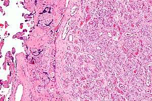 | |
| Micrograph of a chorangioma (right of image). H&E stain. | |
| Classification and external resources | |
| Specialty | obstetrics |
| MeSH | D006391 |
A chorangioma is a non-neoplastic, hamartoma-like growth in the placenta consisting of blood vessels.[1]
Diagnosis
Large chorangiomas are diagnosed by ultrasound or MRI,[1] and confirmed by histologic examination of the placenta.
Histologically, chorangioma consist of abundant vascular channels and may be cellular.
Significance
Most chorangiomas are not clinically significant, i.e. they do not have an adverse effect on placental function.
The significance of a chorangioma is determined by its size and whether it is found together with other chorangiomas. Chorangiomas are significant if multiple or "large", i.e. greater than 4 cm[1] or 5 cm.[2]
Treatment
Small chorangiomas are not treated. Large chorangioma can be treated several ways, including chemical ablation and laser coagulation.[1]
See also
Additional images
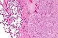 Micrograph of a chorangioma (right of image) and normal placenta (left of image). H&E stain.
References
- 1 2 3 4 Amer HZ, Heller DS (2010). "Chorangioma and related vascular lesions of the placenta--a review". Fetal Pediatr Pathol. 29 (4): 199–206. doi:10.3109/15513815.2010.487009. PMID 20594143.
- ↑ Lež C, Fures R, Hrgovic Z, Belina S, Fajdic J, Münstedt K (2010). "Chorangioma placentae". Rare Tumors. 2 (4): e67. doi:10.4081/rt.2010.e67. PMC 3019602. PMID 21234259.