Central canal
| Central canal of spinal cord | |
|---|---|
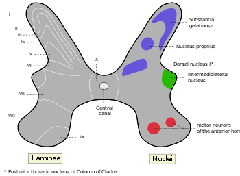
 Cross-section through cervical spinal cord. | |
 | |
| Details | |
| Identifiers | |
| Latin | canalis centralis medullae spinalis |
| TA | A14.1.02.019 |
| FMA | 78497 |
| Anatomical terminology | |
The central canal, also known as ependymal canal, is the cerebrospinal fluid-filled space that runs longitudinally through the length of the entire spinal cord. The central canal is continuous with the ventricular system of the brain. The fourth ventricle narrows at a region called the obex to become the central canal of the spinal cord. The central canal helps to transport nutrients to the spinal cord as well as protect it by cushioning the impact of a force when the spine is affected
The central canal represents the adult remainder of the central cavity of the neural tube. It generally occludes (closes off) with age.[1]
Structure
Clinical significance
Syringomyelia is a disease caused by the occlusion of the central canal. Occlusions of the central canal typically occur at the lower cervical and upper thoracic levels. This typically damages white matter fibers that cross in anterior white commissure, leading to the loss of temperature, pain, and motor function at the affected levels on contralateral sides.
Terminal ventricle
The terminal ventricle (ventriculus terminalis, fifth ventricle or ampulla caudalis) is the widest part of the central canal of the spinal cord that is located at or near the conus medullaris.[2] It was described by Stilling in 1859 and Krause in 1875.[3] Krause introduced the term fifth ventricle after observation of normal ependymal cells.[3] The central canal expands as a fusiform terminal ventricle, and approximately 8–10 mm in length in the conus medullaris (or conus terminalis).[4] Although the terminal ventricle is visible in the fetus and children, it is usually absent in adults.[2]
Sometimes, the terminal ventricle is observed by MRI or ultrasound in children less than 5 years old.[5] An MRI scan can be particularly helpful in its detection.
In pathological conditions, an MRI is useful at the level of the conus medullaris. Findings may be related to the following:
- Spina bifida
- Arnold-Chiari syndrome
- Tumors of the conus medullaris
- Myelomeningocele
- Syringomyelia
- Hydromyelia. In hydromyelia, a dilation of the central canal of the spinal cord is caused by an increase of cerebrospinal fluid.[6]
- Syringohydromyelia (i.e., both Syringomyelia and Hydromyelia)[6]
- Tethered cord
In some cases, the terminal ventricle may cause clinical symptoms due to its expansion.
Central gelatinous substance
| Central gelatinous substance of spinal cord | |
|---|---|
|
Substantia gelatinosa centralis is Rexed lamina X, labeled at center. | |
| Details | |
| Identifiers | |
| Latin | substantia gelatinosa centralis medullae spinalis |
| TA | A14.1.02.019 |
| FMA | 78497 |
| Anatomical terminology | |
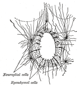
Throughout the cervical and thoracic regions the central canal is situated in the anterior third of the spinal cord; in the lumbar enlargement it is near the middle, and in the conus medullaris it approaches the posterior surface. It is filled with cerebrospinal fluid, and lined by ciliated, columnar epithelium, outside of which is an encircling band of gelatinous substance, the substantia gelatinosa centralis (or central gelatinous substance of spinal cord). This gelatinous substance consists mainly of neuroglia, but contains a few nerve cells and fibers; it is traversed by processes from the deep ends of the columnar ciliated cells which line the central canal.
The substantia gelatinosa of Rolando, is located more dorsally.
See also
References
- ↑ Yasui K, Hashizume Y, Yoshida M, Kameyama T, Sobue G (1999). "Age-related morphologic changes of the central canal of the human spinal cord". Acta Neuropathol. 97 (3): 253–9. doi:10.1007/s004010050982. PMID 10090672.
- 1 2 "ventriculus terminalis". radsource.us. July 2008.
- 1 2 Liccardo G, Ruggeri F, De Cerchio L, Floris R, Lunardi P (June 2005). "Fifth ventricle: an unusual cystic lesion of the conus medullaris". Spinal Cord. 43 (6): 381–4. doi:10.1038/sj.sc.3101712. PMID 15655569.
- ↑ Williams & Warwick. Gray's Anatomy .THIRTY-SEVENTH EDITION. ISBN 0 443 04177 6
- ↑ Celli P, D'Andrea G, Trillò G, Roperto R, Acqui M, Ferrante L (March 2002). "Cyst of the medullary conus: malformative persistence of terminal ventricle or compressive dilatation?". Neurosurgical Review. 25 (1–2): 103–6. doi:10.1007/s10143-001-0203-8. PMID 11954762.
- 1 2 "imaging in syringohydromyelia". emedicine.medscape. 2018-04-25.
This article incorporates text in the public domain from page 753 of the 20th edition of Gray's Anatomy (1918)
External links
- "Anatomy diagram: 13048.000-3". Roche Lexicon - illustrated navigator. Elsevier. Archived from the original on 2014-01-01.
- ancil-993 at NeuroNames