Breast MRI
| Breast MRI | |
|---|---|
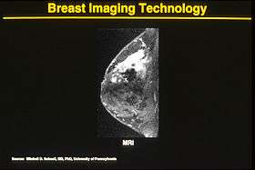 Breast MRI demonstrating marked enhancement (bright area) which was confirmed to be cancer. | |
| ICD-10-PCS | BH3 |
| ICD-9-CM | 88.92, 88.97 |
| OPS-301 code | 3-827 |
One alternative to mammography, Breast MRI or contrast enhanced magnetic resonance imaging (MRI), has shown substantial progress in the detection of breast cancer.
Indications[1]
- Screening for malignancy in women with greater than 20% lifetime risk of breast cancer.
- Evaluate breast implants for rupture.
- Screening the contralateral breast for malignancy in women with known unilateral breast malignancy.
- Extent of disease and the presence of multifocality and multicentricity in patients with invasive carcinoma and ductal carcinoma in situ (DCIS).
- Evaluate response to neoadjuvant chemotherapy.
Comparison to Other Technologies
The available literature suggests that the sensitivity of contrast-enhanced breast MRI in detection of cancer is considerably higher than that of either radiographic mammography or ultrasound and is generally reported to be in excess of 94%.[2]
The specificity (the confidence that a lesion is cancerous and not a false positive) is only fair ('modest'),[3] (or 37%-97%[2]) thus a positive finding by MRI should not be interpreted as a definitive diagnosis.
The reports of 4,271 breast MRIs from eight large scale clinical trials were reviewed in 2006.[4]
Nephrogenic Systemic Fibrosis (NSF)
This systemic disease resembles scleromyxedema and to some extent scleroderma. It may occur months after contrast has been injected.[11] Patients with poorer renal function are more at risk for NSF, with dialysis patients being more at risk than patients with renal insufficiency.[12][13] After several years of controversy during which up to 100 Danish patients have been gadolinium poisoned (and some died) after use of the contrast agent Omniscan, it was admitted by the Norwegian medical company Nycomed that they were aware of some dangers of using gadolinium-based agents for their product.[14] At present, NSF has been linked to the use of four gadolinium-containing MRI contrast agents.[5]
See also
References
- ↑ https://www.acr.org/~/media/2a0eb28eb59041e2825179afb72ef624.pdf
- 1 2 Magnetic Resonance Imaging of the Breast
- ↑ DeMartini W, Lehman C, Partridge S (April 2008). "Breast MRI for cancer detection and characterization: a review of evidence-based clinical applications". Acad Radiol. 15 (4): 408–16. doi:10.1016/j.acra.2007.11.006. PMID 18342764.
- ↑ Lehman CD (2006). "Role of MRI in screening women at high risk for breast cancer". Journal of Magnetic Resonance Imaging. 24 (5): 964–70. doi:10.1002/jmri.20752. PMID 17036340.
- ↑ MRI contrast agent