Basal lamina
| Basal lamina | |
|---|---|
 | |
| Details | |
| Identifiers | |
| Latin | lamina basalis |
| MeSH | D016570, D001485 |
| TA |
A15.2.03.007 A15.2.03.019 A15.3.03.102 |
| TH | H2.00.00.0.00006 |
| FMA | 62918 58438, 62918 |
|
Anatomical terms of microanatomy | |
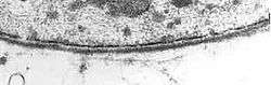
The basal lamina is a layer of extracellular matrix secreted by the epithelial cells, on which the epithelium sits. It is often incorrectly referred to as the basement membrane, though it does constitute a portion of the basement membrane. The basal lamina is visible only with the electron microscope, where it appears as an electron-dense layer that is 20–100 nm thick (with some exceptions that are thicker, such as basal lamina in lung alveoli and renal glomeruli).
Structure
The layers of the basal lamina ("BL") and those of the basement membrane ("BM") are described below:
| Name | Part of BL? | Part of BM? | Notes |
| lamina lucida / lamina rara interna[1] | yes | yes | electron-lucid layer[2] containing the glycoprotein laminin |
| lamina densa | yes | yes | electron-dense layer[3] composed of type IV collagen |
| lamina lucida / lamina rara externa | yes | yes | Similar composition to lamina rara interna. Some sources do not consider this a distinct layer. |
| lamina reticularis[4] | no | yes | The three above layers of the basal lamina typically sit on top of the reticular lamina, which is synthesized by cells from the underlying connective tissue and contains fibronectin. The exception is when two epithelial layers abut one another as in the alveoli of the lungs and glomeruli of the kidneys, in which the basal lamina of one epithelial layer fuses with that of the other. |
Anchoring fibrils composed of type VII collagen extend from the basal lamina into the underlying reticular lamina and loop around collagen bundles. Although found beneath all basal laminae, they are especially numerous in stratified squamous cells of the skin.
These layers should not be confused with the lamina propria, which is found outside the basal lamina.[5]
Basement membrane
The basement membrane is visible under light microscopy. Electron microscopy shows that the basement membrane consists of three layers: the lamina lucida (electron-lucent), lamina densa (electron-dense), and lamina fibroreticularis (electron-lucent).
The lamina densa was formerly known as the basal lamina. The terms basal lamina and basement membrane were often used interchangeably, until it was realised that all three layers seen with the electron microscope represent the single layer seen with the light microscope. This has led to considerable terminological confusion and, if used, the term basal lamina should be confined to its meaning as lamina densa.[6]
Some theorize that the lamina lucida is an artifact created when preparing the tissue, and that the lamina lucida is therefore equal to the lamina densa in vivo.[7]
The term "basal lamina" is usually used with electron microscopy, while the term "basement membrane" is usually used with light microscopy.
Examples of basement membranes include:
Function
The basal lamina is made and maintained by the cells that sit on it. It acts as a point of attachment for cells. However, it can also have other function such as a permeability barrier in the glomerulus (urine production). Some of the matrix molecules (of the basal lamina) mediate synaptic adhesion in neuromuscular synapses (citation: [edited by] Bradley G. Klein. Cunningham's Textbook of Veterinary Physiology. St. Louis, Mo. :Elsevier/Saunders, 2013. Print.-page61).
Alport syndrome is a genetic disorder resulting from mutations in the COL4A3/4/5 genes. These genes are important in collagen IV synthesis and basement membrane formation. In individuals with this syndrome the basement membrane in structures such as the glomerulus, ears, and eyes does not function properly, causing symptoms such as blood in the urine, loss of hearing, and vision problems.
See also
References
- ↑ Histology image:22403loa from Vaughan, Deborah (2002). A Learning System in Histology: CD-ROM and Guide. Oxford University Press. ISBN 978-0195151732.
- ↑ UIUC Histology Subject 500
- ↑ UIUC Histology Subject 499
- ↑ Histology image:20904loa from Vaughan, Deborah (2002). A Learning System in Histology: CD-ROM and Guide. Oxford University Press. ISBN 978-0195151732.
- ↑ Histology image:22203loa from Vaughan, Deborah (2002). A Learning System in Histology: CD-ROM and Guide. Oxford University Press. ISBN 978-0195151732.
- ↑ Wheater's Functional Histology 4th edition (2000)
- ↑ Chan F, Inoue S (1994). "Lamina lucida of basement membrane: an artefact". Microsc Res Tech. 28 (1): 48–59. doi:10.1002/jemt.1070280106. PMID 8061357.