Abdominal wall
| Abdominal wall | |
|---|---|
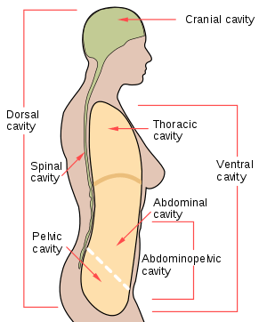 Body cavities | |
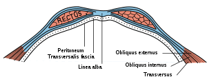 Diagram of sheath of Rectus above the arcuate line. | |
| Details | |
| Identifiers | |
| Latin | paries abdominalis |
| MeSH | D034861 |
| FMA | 299989 |
| Anatomical terminology | |
In anatomy, the abdominal wall represents the boundaries of the abdominal cavity. The abdominal wall is split into the posterior (back), lateral (sides) and anterior (front) walls.
There is a common set of layers covering and forming all the walls: the deepest being the visceral peritoneum, which covers many of the abdominal organs (most of the large and small intestines, for example), and the parietal peritoneum- which covers the visceral peritoneum below it, the extraperitoneal fat, the transversalis fascia, the internal and external oblique and transversus abdominis aponeurosis, and a layer of fascia, which has different names according to what it covers (e.g., transversalis, psoas fascia).
In medical vernacular, the term 'abdominal wall' most commonly refers to the layers composing the anterior abdominal wall which, in addition to the layers mentioned above, includes the three layers of muscle: the transversus abdominis (transverse abdominal muscle), the internal (obliquus internus) and the external oblique (obliquus externus).
Layers of anterior abdominal wall
In human anatomy, the layers of the abdominal wall are (from superficial to deep):
- Skin
- Subcutaneous tissue
- Fascia
- Camper's fascia - fatty superficial layer.
- Scarpa's fascia - deep fibrous layer.
- Muscle
- Fascia transversalis
- Peritoneum
Inner surface
The surface contains several ligaments separated by fossae:
| Ligament/fold | Remnant of | Lateral fossa | Hernia |
| median umbilical ligament | urachus | supravesical fossa | supravesical hernia (rare) |
| medial umbilical ligament | umbilical artery | medial inguinal fossa | direct inguinal hernia |
| lateral umbilical fold | inferior epigastric vessels | lateral inguinal fossa | indirect inguinal hernia |
See also
External links
- skel&wallsabd at The Anatomy Lesson by Wesley Norman (Georgetown University) - "Skeleton of the Abdomen", Wesley Norman, PhD, DSc
- Abdominal+Wall at the US National Library of Medicine Medical Subject Headings (MeSH)
- Anterolateral Abdominal Wall - University of Edinburgh Faculty of Medicine
- Muscles of the Anterior Abdominal Wall - University of Arkansas