Venography
Venography (also called phlebography or ascending phlebography) is a procedure in which an x-ray of the veins, a venogram, is taken after a special dye is injected into the bone marrow or veins. The dye has to be injected constantly via a catheter, making it an invasive procedure. Normally the catheter is inserted by the groin and moved to the appropriate site by navigating through the vascular system.
| Venography | |
|---|---|
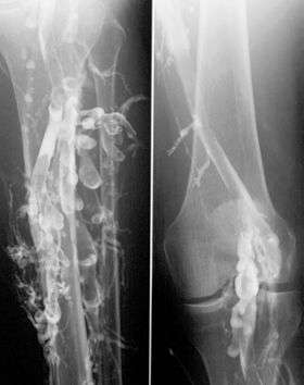 Phlebography in a patient with deep venous thrombosis | |
| ICD-9-CM | 88.6 |
| MeSH | D010690 |
| OPS-301 code | 3-61 |
| MedlinePlus | 007283 |
Contrast venography is the gold standard for judging diagnostic imaging methods for deep venous thrombosis; although, because of its cost, invasiveness, and other limitations this test is rarely performed.[1]
Venography can also be used to distinguish blood clots from obstructions in the veins, to evaluate congenital vein problems, to see how the deep leg vein valves are working, or to identify a vein for arterial bypass grafting.
Areas of the venous system that can be investigated include the lower extremities, the inferior vena cava, and the upper extremities.
The United States National Institute of Health says the following about varicose veins, "they cause concern and distress on a large scale, most of which can be dealt with by good explanation and reassurance, or by a variety of treatments which are evolving rapidly at present." [2]
See also
References
- Bates SM, Jaeschke R, Stevens SM, et al. (2012). "Diagnosis of DVT: Antithrombotic Therapy and Prevention of Thrombosis, 9th ed: American College of Chest Physicians Evidence-Based Clinical Practice Guidelines". Chest. 141 (suppl 2): e383S. doi:10.1378/chest.11-2299. PMC 3278048. PMID 22315267.
- "Post | Varicose Vein & Spider Vein Treatment | Dallas Vein Institute". dallasveininstitute. 2019-07-11. Retrieved 2019-07-12.
External links
- Baylor College of Medicine Venogram procedure page, retrieved November 4, 2006
- WebMD Venogram procedure page, retrieved November 4, 2006
| X-ray/ Radiography |
| ||||||||||||
|---|---|---|---|---|---|---|---|---|---|---|---|---|---|
| MRI | |||||||||||||
| Ultrasound | |||||||||||||
| Radionuclide |
| ||||||||||||
| Optical/Laser | |||||||||||||
| Thermography |
| ||||||||||||
| Target conditions |
| ||||||||||||
| |||||||||||||