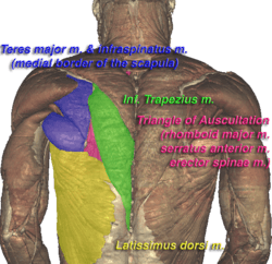
Triangle of auscultation
The triangle of auscultation of the lungs is a relative thinning of the musculature of the back, situated along the medial border of the scapula.
| Triangle of auscultation | |
|---|---|
 Triangle of auscultation (shown in pink) of the Visible Human Male, created in the VH Dissector | |
 Cross section #1428 of the Visible Human Male showing the structures of the triangle of auscultation, created in the VH Dissector | |
| Details | |
| Identifiers | |
| Latin | trigonum auscultationis |
| TA | A01.2.05.006 |
| FMA | 75022 |
| Anatomical terminology | |
Boundaries
It has the following boundaries:
- Superiorly and medially, by the inferior portion of the trapezius
- Inferiorly, by the latissimus dorsi
- Laterally, by the medial border of the scapula
The superficial floor of the triangle is formed by the lateral portion of the erector spinae muscles. Deep to these muscles are the osseous portions of the 6th and 7th ribs and the internal and external intercostal muscles.
Function
Due to the relative thinning of the musculature of the back in the triangle, the posterior thoracic wall is closer to the skin surface, making respiratory sounds able to be heard more clearly with a stethoscope.
Typically, the triangle of auscultation is covered by the scapula. To better expose the floor of the triangle up of the posterior thoracic wall in the 6th and 7th intercostal space, a patient is asked to fold their arms across their chest, laterally rotating the scapulae, while bending forward at the trunk, somewhat resembling a fetal position.
On the left side, the cardiac orifice of the stomach lies deep to the triangle, and in days before X-rays were discovered the sound of swallowed liquids were auscultated over this triangle to confirm an oesophageal tumour
References
This article incorporates text in the public domain from page 434 of the 20th edition of Gray's Anatomy (1918)
External links
- Anatomy photo:01:02-0103 at the SUNY Downstate Medical Center
- Overview and diagram at ithaca.edu
| Authority control |
|---|