Somatopleuric mesenchyme
In the anatomy of an embryo, the somatopleue is a structure created during embryogenesis when the lateral mesoderm splits into two layers. The outer (or somatic) layer becomes applied to the inner surface of the ectoderm, and with it forms the somatopleure.[1]
| Somatopleuric mesenchyme | |
|---|---|
 A series of a transverse sections through an embryo of the dog. (After Bonnet.) Section I is the most anterior. In V the neural plate is spread out nearly flat. The series shows the uprising of the neural folds to form the neural canal. a. Aortæ. c. Intermediate cell mass. ect. Ectoderm. ent. Entoderm. h, h. Rudiments of endothelial heart tubes. In III, IV, and V the scattered cells represented between the entoderm and splanchnic layer of mesoderm are the vasoformative cells which give origin in front, according to Bonnet, to the heart tubes, h; l.p. Lateral plate still undivided in I, II, and III; in IV and V split into somatic (sm) and splanchnic (sp) layers of mesoderm. mes. Mesoderm. p. Pericardium. so. Primitive segment. | |
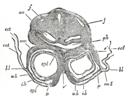 Transverse section through the region of the heart in a rabbit embryo of nine days. X 80. (Kölliker.) j, j. Jugular veins. ao. Aorta. ph. Pharynx. som. Somatopleure. bl. Proamnion. ect. Ectoderm. ent. Entoderm. p. Pericardium. spl. Splanchnopleure. ah. Outer wall of heart. ih. Endothelial lining of heart. é. Septum between heart tubes. | |
| Details | |
| Carnegie stage | 9 |
| Precursor | lateral plate mesoderm, ectoderm |
| Gives rise to | mesenchyme |
| Identifiers | |
| Latin | mesenchyma somatopleurale |
| TE | E4.0.4.1.0.0.3 |
| Anatomical terminology | |
The combination of ectoderm and mesoderm, or somatopleure, forms the amnion, the chorion and the lateral body wall of the embryo. The limbs are formed from the somatic mesoderm cells are induced by hox genes and the expression of other molecules to suffer and epithelial-mesenchyme transition.[1] The embryionic somatopleure is then divided into 3 sections, the anterior limb bud formation, the posterior limb bud formation and the non limb forming wall. The bud forming sections grow in size at the somatic mesoderm under the ectoderm proliferate in mesenchyme form.[1]
In chicken, the extraembryonic tissues are separated into two layers: the splanchnopleure composed of the endoderm and splanchnic mesoderm, and the somatopleure composed of the ectoderm and somatic mesoderm along with the formation of the coelomic cavity after gastrulation. The amnion and chorion are derived from the somatopleure with a presumptive border of the ectamnion.[2] Following the anterior extension of the extraembryonic mesoderm and formation of the coelom, the anterior and lateral amniotic folds arise along the ectamnion and grow posteriorly over the head of the embryo.[2] A portion of the amniogenic somatopleure adjacent to the base of the head fold is identified as the region contributing to embryonic tissues in the thoracic wall and pharyngeal and cardiac regions. The somatopleure is known to serve as the matrix of the ventrolateral body wall and gives rise to connective tissue, tendons and the sternum.[2][3]
See also
References
This article incorporates text in the public domain from page 50 of the 20th edition of Gray's Anatomy (1918)
- Gilbert, Scott F., 1949- (2014). Developmental biology (Tenth ed.). Sunderland, MA, USA. ISBN 978-0-87893-978-7. OCLC 837923468.CS1 maint: multiple names: authors list (link)
- Asai, Rieko; Haneda, Yuka; Seya, Daiki; Arima, Yuichiro; Fukuda, Kimiko; Kurihara, Yukiko; Miyagawa-Tomita, Sachiko; Kurihara, Hiroki (2017-08-21). "Amniogenic somatopleure: a novel origin of multiple cell lineages contributing to the cardiovascular system". Scientific Reports. 7 (1): 8955. Bibcode:2017NatSR...7.8955A. doi:10.1038/s41598-017-08305-2. ISSN 2045-2322. PMID 28827655.

- Christ, Bodo; Huang, Ruijin; Scaal, Martin (2007). "Amniote somite derivatives". Developmental Dynamics. 236 (9): 2382–2396. doi:10.1002/dvdy.21189. ISSN 1058-8388. PMID 17557304.
External links
- Diagram at Yuba Community College District at the Library of Congress Web Archives (archived 2008-08-09)
- Overview at Kennesaw State University
| Authority control |
|---|