Pinocytosis
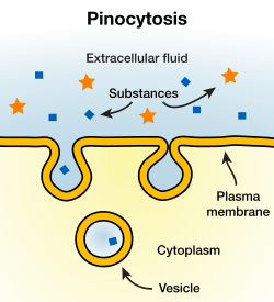
In cellular biology, pinocytosis, otherwise known as fluid endocytosis and bulk-phase pinocytosis, is a mode of endocytosis in which small particles suspended in extracellular fluid are brought into the cell through an invagination of the cell membrane, resulting in a suspension of the particles within a small vesicle inside the cell. These pinocytotic vesicles subsequently fuse with endosomes to hydrolyze (break down) the particles.
Pinocytosis is further segregated into the pathways macropinocytosis, clathrin-mediated endocytosis, caveolin-mediated endocytosis, or clathrin- and caveolin-independent endocytosis, all of which differ by the mechanism of vesicle formation as well as the resulting size of these vesicles.
Pinocytosis is variably subdivided into categories depending on molecular mechanism and the fate of the internalized molecules. Pinocytosis is, in some cases, considered to be a constitutive process, while in others it is receptor-mediated and highly regulated. One scheme divides pinocytosis into the four categories of caveolae-mediated, clathrin-dependent, macropinocytosis, and dynamin and clathrin-independent Seto et al (2002).
Etymology and pronunciation
The word pinocytosis (/ˌpɪnəsaɪˈtoʊsɪs, ˌpaɪ-, -noʊ-, -sə-/[2][3][4]) uses combining forms of pino- + cyto- + -osis, all New Latin from Greek, reflecting píno, to drink, and cytosis. The term was proposed by W. H. Lewis in 1931.[5]
Non-specific, adsorptive pinocytosis
Non-specific, adsorptive pinocytosis is a form of endocytosis, a process in which small particles are taken in by a cell by splitting off small vesicles from the cell surface.[6] Cationic proteins bind to the negative cell surface and are taken up via the clathrin-mediated system, thus the uptake is intermediate between receptor-mediated endocytosis and non-specific, non-adsorptive pinocytosis. The clathrin-coated pits occupy about 2% of the surface area of the cell and only last about a minute, with an estimated 2500 leaving the average cell surface each minute. The clathrin coats are lost almost immediately, and the membrane is subsequently recycled to the cell surface.
References
- Abbas, Abul, et al. "Basic Immunology: Functions and Disorders of the Immune System." 5th ed. Elsevier, 2016. p.69
- "Pinocytosis". Oxford Dictionaries UK Dictionary. Oxford University Press. Retrieved 2016-01-22.
- "Pinocytosis". Merriam-Webster Dictionary. Retrieved 2016-01-22.
- "Pinocytosis". Dictionary.com Unabridged. Random House. Retrieved 2016-01-22.
- Rieger, R.; Michaelis, A.; Green, M.M. 1991. Glossary of Genetics. Classical and Molecular (Fifth edition). Springer-Verlag, Berlin, .
- Alberts, Johnson, Lewis, Raff, Roberts, Walter: "Molecular Biology of the Cell", Fourth Edition, Copyright 2002 P.748
- Campbell, Reece, Mitchell: "Biology", Sixth Edition, Copyright 2002 P. 151
- Marshall, Ben, Incredible Biological Advancements of the 20th Century, Copyright 2001 p. 899
- Alrt, Pablo, Global Society Harvard study, copyright 2003 p. 189
- Brooker, Robert: "Biology", Second Edition, Copyright 2011 p. 116
- Cherrr, Malik, The Only Edition, Copyright 2012, p. 256
- Abbas, Abul, et al. "Basic Immunology: Functions and Disorders of the Immune System." 5th ed. Elsevier, 2016. p.69