Nerve of pterygoid canal
The nerve of the pterygoid canal (Vidian nerve) is formed by the junction of the greater petrosal nerve and deep petrosal nerve, which passes from the foramen lacerum to the pterygopalatine fossa through the pterygoid canal.
| Nerve of pterygoid canal | |
|---|---|
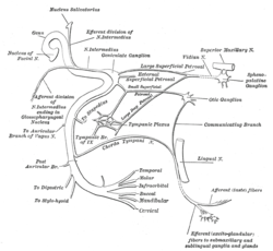 Plan of the facial and intermediate nerves and their communication with other nerves. (Vidian nerve labeled at upper right.) | |
| Details | |
| From | greater petrosal nerve, deep petrosal nerve |
| To | pterygopalatine ganglion |
| Identifiers | |
| Latin | n. canalis pterygoidei |
| TA | A14.3.02.007 |
| FMA | 67584 |
| Anatomical terms of neuroanatomy | |
Structure
The nerve of the pterygoid canal forms from the junction of the greater petrosal nerve and the lesser petrosal nerve within the foreamen lacerum. This combined nerve exits the foramen lacerum and travels to the pterygopalatine fossa through the pterygoid canal in the sphenoid.
The nerve of the pterygoid canal contains axons of both sympathetic and parasympathetic axons, specifically;
- preganglonic parasympathetic axons from the greater petrosal nerve, a branch of the facial nerve (cell bodies are located in the superior salivatory nucleus)
- postganglionic sympathetic axons from the deep petrosal nerve, a branch of the internal carotid plexus (cell bodies are located in the superior cervical ganglion)
Function
The preganglionic parasympathetic axons synapse in the pterygopalatine ganglion, which contains the postganglionic neurons which provide secretomotor innervation to the lacrimal gland, as well as the nasal and palatine glands.
The postganglionic sympathetic axons do not synapse in the pterygopalatine ganglion, they travel on the branches of the maxillary nerve to provide sympathetic innervation to blood vessels.
Additional images
 Alveolar branches of superior maxillary nerve and pterygopalatine ganglion.
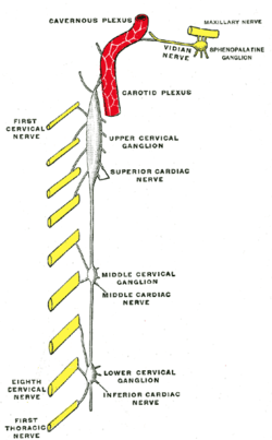 Diagram of the cervical sympathetic.
See also
References
This article incorporates text in the public domain from page 892 of the 20th edition of Gray's Anatomy (1918)
External links
- cranialnerves at The Anatomy Lesson by Wesley Norman (Georgetown University) (VII) ("NPC")
Anatomy of the autonomic nervous system | |||||
|---|---|---|---|---|---|
| Head |
| ||||
| Neck |
| ||||
| Chest |
| ||||
| Abdomen |
| ||||
| Pelvis |
| ||||
| |||||
| Authority control |
|---|