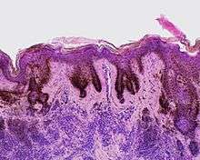
Liver spot
Liver spots (also known as age spot, solar lentigo,[1] "lentigo senilis",[1]:686 "old age spot",[2] "senile freckle"[2]) are blemishes on the skin associated with aging and exposure to ultraviolet radiation from the sun. They range in color from light brown to red or black and are located in areas most often exposed to the sun, particularly the hands, face, shoulders, arms and forehead, and the scalp if bald.
| Liver spot | |
|---|---|
 | |
| Liver spots (solar lentigo) on the left hand of a 63-year-old light-skinned Caucasian man | |
| Specialty | Dermatology |
The spots derive their name from the fact that they were once incorrectly believed to be caused by liver problems, but they are physiologically unrelated to the liver, save for a similar colour.[3] From the age of 40 onward the skin is less able to regenerate from sun exposure, and liver spots are very common in this age group, particularly in those who spend time in the sun.
In the overwhelming majority of cases, liver spots pose no threat and require no treatment, though they occasionally have been known to obscure the detection of skin cancer. However, despite being a benign condition, liver spots are sometimes considered unsightly and some people choose to have them removed. This can be done by electrosurgery, laser treatment, cryotherapy, or the use of depigmentation agents, such as hydroquinone,[4] tretinoin,[4] topical cysteamine,[5] azelaic acid,[6] or alpha hydroxy acids.[7]
Causes
Differently from the melanotic nevi and the verrucous nevi on the skin, age spots change in color and in shape with time. Michelitsch and Michelitsch propose an hypothesis inspired by their misrepair-accumulation aging theory[8][9] for the development of age spots.[10] They propose that aged basal cells contain lipofuscin bodies cannot be removed and might promote the aging of neighboring, generating a feedback loop whereby more and more neighbor cells become aged and lipofuscin-containing.[10] Such cells might then aggregate into a spot with an irregular shape.[10] They propose that the protruding of a flat spot is a result of the death of aged cells in the spot and release of lipofuscin bodies.[10] The aggregating cells would form a capsule, and the dense lipofuscin bodies make the protruding spot soft and dark in color.[10] However, this proposal appeared as a preprint in 2015, has little direct empirical support, and has never been published in a peer reviewed journal.
Another group[11] has reported that "age spots" taken from human skin biopsies of patients facial senile lentigo of Fitzpatrick skin type III or IV aged 55-62 are enriched with senescent fibroblasts compared to surrounding skin. The dark coloration appeared to be due to higher melanin levels and activity of tyrosinase in the senescent fibroblasts than in the controls, potentially related to lower SDF1 expression.[11] Patients were then administered six weekly treatments of microneedle fractional radiofrequency aimed at eliminating dermal senescent fibroblasts; this led to a marked decrease in epidermal pigmentation compared to baseline, accompanied by a decrease in the synthesis of collagen and the normalization of suppressed SDF1 expression.[11]
Diagnosis
The diagnosis is usually done by a dermatologist.
References
- James, William D.; Berger, Timothy G.; et al. (2006). Andrews' Diseases of the Skin: Clinical Dermatology. Saunders Elsevier. ISBN 0-7216-2921-0.
- Rapini, Ronald P.; Bolognia, Jean L.; Jorizzo, Joseph L. (2007). Dermatology: 2-Volume Set. St. Louis: Mosby. pp. 1716–17. ISBN 1-4160-2999-0.
- Karen J. Carlson, Stephanie A. Eisenstat, Terra Diane Ziporyn, The new Harvard guide to women's health, Harvard University Press, 2004, p. 337.
- "Age spots (liver spots) Treatments and drugs". Mayo Clinic. Retrieved 2017-02-12.
- Mansouri, P.; Farshi, S.; Hashemi, Z.; Kasraee, B. (2015-07-01). "Evaluation of the efficacy of cysteamine 5% cream in the treatment of epidermal melasma: a randomized double-blind placebo-controlled trial". The British Journal of Dermatology. 173 (1): 209–17. doi:10.1111/bjd.13424. ISSN 1365-2133. PMID 25251767.
- Rodríguez Prieto, M. A.; Manchado Lopez, P.; Ruiz Gonzalez, I.; Suarez, D. (1993-05-01). "Treatment of lentigo maligna with azelaic acid". International Journal of Dermatology. 32 (5): 363–64. doi:10.1111/j.1365-4362.1993.tb01475.x. ISSN 0011-9059. PMID 8505164.
- "Overview about liver spots/age spots". Beauty Natural Skin. Retrieved 3 July 2015.
- Wang, Jicun; Michelitsch, Thomas; Wunderlin, Arne; Mahadeva, Ravi (2009). "Aging as a consequence of Misrepair – a novel theory of aging". 0904: 9. arXiv:0904.0575. Bibcode:2009arXiv0904.0575W. Cite journal requires
|journal=(help) - Wang-Michelitsch, Jicun; Michelitsch, Thomas (2015). "Aging as a process of accumulation of Misrepairs". 1503: 13. arXiv:1503.07163. Bibcode:2015arXiv150307163W. Cite journal requires
|journal=(help) - Wang-Michelitsch, Jicun; Michelitsch, Thomas (2015). "Development of age spots as a result of accumulation of aged cells in aged skin". 1505: 9. arXiv:1505.07012. Bibcode:2015arXiv150507012W. Cite journal requires
|journal=(help) - Yoon JE, Kim Y, Kwon S, Kim M, Kim YH, Kim JH, Park TJ, Kang HY (September 9, 2018). "Senescent fibroblasts drive ageing pigmentation: A potential therapeutic target for senile lentigo". Theranostics. 8 (17): 4620–4632. doi:10.7150/thno.26975. PMC 6160768. PMID 30279727. Retrieved 26 December 2019.CS1 maint: multiple names: authors list (link)