Large-scale brain networks
Large-scale brain networks are collections of widespread brain regions showing functional connectivity by statistical analysis of the fMRI BOLD signal[1] or other recording methods such as EEG,[2] PET[3] and MEG.[4] An emerging paradigm in neuroscience is that cognitive tasks are performed not by individual brain regions working in isolation but by networks consisting of several discrete brain regions that are said to be "functionally connected" due to tightly coupled activity. Functional connectivity may be measured as long-range synchronization of the EEG, MEG, or other dynamic brain signals.[5] Synchronized brain regions may also be identified using spatial independent component analysis (ICA).
The set of identified brain areas that are linked together in a large-scale network varies with cognitive function.[6] When the cognitive state is not explicit (i.e., the subject is at "rest"), the large-scale brain network is a resting state network (RSN). As a physical system with graph-like properties,[5] a large-scale brain network has both nodes and edges and cannot be identified simply by the co-activation of brain areas. In recent decades, the analysis of brain networks was made feasible by advances in imaging techniques as well as new tools from graph theory and dynamical systems.
Large-scale brain networks are identified by their function and provide a coherent framework for understanding cognition by offering a neural model of how different cognitive functions emerge when different sets of brain regions join together as self-organized coalitions. The identification of the coalitions will vary with different parameters used to run the ICA algorithm,[7] which can result in a different number of networks. In one model, there is only the default mode network and the task-positive network, but most current analyses show several networks, which are enumerated below. The property called functional network flexibility a brain region with strong functional connections within a brain network suddenly establish many connections to a different network.[8]
Disruptions in activity in various networks have been implicated in neuropsychiatric disorders such as depression, Alzheimer's, autism spectrum disorder, schizophrenia and bipolar disorder.[9]
Networks
The following six networks have been identified by at least three studies.
Default mode
- The default mode network is active when an individual is awake and at rest. It preferentially activates when individuals focus on internally-oriented tasks such as daydreaming, envisioning the future, retrieving memories, and theory of mind. It is negatively correlated with brain systems that focus on external visual signals. It is the most widely researched network.[5][8][10][1][11][12][13][14][15]
Dorsal attention
- This network is involved in the voluntary deployment of attention and reorientation to unexpected events.[1][11][12][14][15][16][17] Within the dorsal attention network, the intraparietal sulcus and frontal eye fields influence the visual areas of the brain. These influencing factors allow for the orientation of attention.[18][16]
Ventral attention
- Three areas of the brain are active in this network, and they include the visual cortex, temporoparietal junction, and the ventral frontal cortex. These areas respond when behaviorally relevant stimuli occur unexpectedly.[16] The ventral attention network may also become inhibited during focused attention in which top down processing is being used, such as when one is visually searching for something. This response may prevent goal driven attention from being distracted by non-relevant stimuli. It becomes active again when the target, or relevant information about the target is found.[16][19]
- [12][16][14][15][17]
Salience
- The salience network consists of several structures, including the anterior (bilateral) insula, dorsal anterior cingulate cortex, and three subcortical structures which are the ventral striatum, substantia nigra/ventral tegmental region.[20][21] It plays the key role of monitoring the salience of external inputs and internal brain events.[1][5][8][11][13][14][15] Specifically it aids in directing attention by identifying important biological and cognitive events.[21]
Fronto-parietal
Lateral visual
Several other brain networks have also been identified: auditory,[11][13] motor,[11] right executive,[11][13] posterior default mode,[11] left fronto-parietal,[12] cerebellar,[12][13] spatial attention,[1][5] attention,[8] language,[5][17] left executive,[13] sensorimotor network,[13] somatomotor,[14][15][8] visual,[14][15][8] temporal,[14][15] limbic,[8][14] visual perception,[17] and visual imagery.[17]
See also
References
- Riedl, Valentin; Utz, Lukas; Castrillón, Gabriel; Grimmer, Timo; Rauschecker, Josef P.; Ploner, Markus; Friston, Karl J.; Drzezga, Alexander; Sorg, Christian (January 12, 2016). "Metabolic connectivity mapping reveals effective connectivity in the resting human brain". PNAS. 113 (2): 428–433. Bibcode:2016PNAS..113..428R. doi:10.1073/pnas.1513752113. PMC 4720331. PMID 26712010.
- Foster, Brett L.; Parvizi, Josef (2012-03-01). "Resting oscillations and cross-frequency coupling in the human posteromedial cortex". NeuroImage. 60 (1): 384–391. doi:10.1016/j.neuroimage.2011.12.019. ISSN 1053-8119. PMC 3596417. PMID 22227048.
- Buckner, Randy L.; Andrews‐Hanna, Jessica R.; Schacter, Daniel L. (2008). "The Brain's Default Network". Annals of the New York Academy of Sciences. 1124 (1): 1–38. Bibcode:2008NYASA1124....1B. doi:10.1196/annals.1440.011. ISSN 1749-6632. PMID 18400922.
- Morris, Peter G.; Smith, Stephen M.; Barnes, Gareth R.; Stephenson, Mary C.; Hale, Joanne R.; Price, Darren; Luckhoo, Henry; Woolrich, Mark; Brookes, Matthew J. (2011-10-04). "Investigating the electrophysiological basis of resting state networks using magnetoencephalography". Proceedings of the National Academy of Sciences. 108 (40): 16783–16788. Bibcode:2011PNAS..10816783B. doi:10.1073/pnas.1112685108. ISSN 0027-8424. PMC 3189080. PMID 21930901.
- Bressler, Steven L.; Menon, Vinod (June 2010). "Large scale brain networks in cognition: emerging methods and principles". Trends in Cognitive Sciences. 14 (6): 233–290. doi:10.1016/j.tics.2010.04.004. PMID 20493761. Retrieved 24 January 2016.
- Bressler, Steven L. (2008). "Neurocognitive networks". Scholarpedia. 3 (2): 1567. Bibcode:2008SchpJ...3.1567B. doi:10.4249/scholarpedia.1567.
- Abou Elseoud, Ahmed; Littow, Harri; Remes, Jukka; Starck, Tuomo; Nikkinen, Juha; Nissilä, Juuso; Timonen, Markku; Tervonen, Osmo; Kiviniemi1, Vesa (2011-06-03). "Group-ICA Model Order Highlights Patterns of Functional Brain Connectivity". Frontiers in Systems Neuroscience. 5: 37. doi:10.3389/fnsys.2011.00037. PMC 3109774. PMID 21687724.
- Bassett, Daniella; Bertolero, Max (July 2019). "How Matter Becomes Mind". Scientific American. 321 (1): 32. Retrieved 23 June 2019.
- Menon, Vinod (2011-09-09). "Large-scale brain networks and psychopathology: A unifying triple network model". Trends in Cognitive Sciences. 15 (10): 483–506. doi:10.1016/j.tics.2011.08.003. PMID 21908230.
- Buckner, Randy L. (2012-08-15). "The serendipitous discovery of the brain's default network". NeuroImage. 62 (2): 1137–1145. doi:10.1016/j.neuroimage.2011.10.035. ISSN 1053-8119. PMID 22037421.
- Yuan, Rui; Di, Xin; Taylor, Paul A.; Gohel, Suril; Tsai, Yuan-Hsiung; Biswal, Bharat B. (30 April 2015). "Functional topography of the thalamocortical system in human". Brain Structure and Function. 221 (4): 1971–1984. doi:10.1007/s00429-015-1018-7. PMC 6363530. PMID 25924563.
- Bell, Peter T.; Shine, James M. (2015-11-09). "Estimating Large-Scale Network Convergence in the Human Functional Connectome". Brain Connectivity. 5 (9): 565–74. doi:10.1089/brain.2015.0348. PMID 26005099.
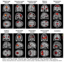
- Heine, Lizette; Soddu, Andrea; Gomez, Francisco; Vanhaudenhuyse, Audrey; Tshibanda, Luaba; Thonnard, Marie; Charland-Verville, Vanessa; Kirsch, Murielle; Laureys, Steven; Demertzi, Athena (2012). "Resting state networks and consciousness. Alterations of multiple resting state network connectivity in physiological, pharmacological and pathological consciousness states". Frontiers in Psychology. 3: 295. doi:10.3389/fpsyg.2012.00295. PMC 3427917. PMID 22969735.
- Yeo, B. T. Thomas; Krienen, Fenna M.; Sepulcre, Jorge; Sabuncu, Mert R.; Lashkari, Danial; Hollinshead, Marisa; Roffman, Joshua L.; Smoller, Jordan W.; Zöllei, Lilla; Polimeni, Jonathan R.; Fischl, Bruce; Liu, Hesheng; Buckner, Randy L. (2011-09-01). "The organization of the human cerebral cortex estimated by intrinsic functional connectivity". Journal of Neurophysiology. 106 (3): 1125–1165. Bibcode:2011NatSD...2E0031H. doi:10.1152/jn.00338.2011. PMC 3174820. PMID 21653723.
- Shafiei, Golia; Zeighami, Yashar; Clark, Crystal A.; Coull, Jennifer T.; Nagano-Saito, Atsuko; Leyton, Marco; Dagher, Alain; Mišić, Bratislav (2018-10-01). "Dopamine Signaling Modulates the Stability and Integration of Intrinsic Brain Networks". Cerebral Cortex. 29 (1): 397–409. doi:10.1093/cercor/bhy264. PMC 6294404. PMID 30357316.
- Vossel, Simone; Geng, Joy J.; Fink, Gereon R. (2014). "Dorsal and Ventral Attention Systems: Distinct Neural Circuits but Collaborative Roles". The Neuroscientist. 20 (2): 150–159. doi:10.1177/1073858413494269. PMC 4107817. PMID 23835449.
- Hutton, John S.; Dudley, Jonathan; Horowitz-Kraus, Tzipi; DeWitt, Tom; Holland, Scott K. (1 September 2019). "Functional Connectivity of Attention, Visual, and Language Networks During Audio, Illustrated, and Animated Stories in Preschool-Age Children". Brain Connectivity. 9 (7): 580–592. doi:10.1089/brain.2019.0679. PMC 6775495. PMID 31144523.
- Fox, Michael D.; Corbetta, Maurizio; Snyder, Abraham Z.; Vincent, Justin L.; Raichle, Marcus E. (2006-06-27). "Spontaneous neuronal activity distinguishes human dorsal and ventral attention systems". Proceedings of the National Academy of Sciences. 103 (26): 10046–10051. Bibcode:2006PNAS..10310046F. doi:10.1073/pnas.0604187103. ISSN 0027-8424. PMC 1480402. PMID 16788060.
- Shulman, Gordon L.; McAvoy, Mark P.; Cowan, Melanie C.; Astafiev, Serguei V.; Tansy, Aaron P.; d'Avossa, Giovanni; Corbetta, Maurizio (2003-11-01). "Quantitative Analysis of Attention and Detection Signals During Visual Search". Journal of Neurophysiology. 90 (5): 3384–3397. doi:10.1152/jn.00343.2003. ISSN 0022-3077. PMID 12917383.
- Steimke, Rosa; Nomi, Jason S.; Calhoun, Vince D.; Stelzel, Christine; Paschke, Lena M.; Gaschler, Robert; Goschke, Thomas; Walter, Henrik; Uddin, Lucina Q. (2017-12-01). "Salience network dynamics underlying successful resistance of temptation". Social Cognitive and Affective Neuroscience. 12 (12): 1928–1939. doi:10.1093/scan/nsx123. ISSN 1749-5016. PMID 29048582.
- Menon, V. (2015-01-01), "Salience Network", in Toga, Arthur W. (ed.), Brain Mapping, Academic Press, pp. 597–611, doi:10.1016/B978-0-12-397025-1.00052-X, ISBN 978-0-12-397316-0, retrieved 2019-12-08
- Scolari, Miranda; Seidl-Rathkopf, Katharina N; Kastner, Sabine (2015-02-01). "Functions of the human frontoparietal attention network: Evidence from neuroimaging". Current Opinion in Behavioral Sciences. Cognitive control. 1: 32–39. doi:10.1016/j.cobeha.2014.08.003. ISSN 2352-1546. PMC 4936532. PMID 27398396.
- Marek, Scott; Dosenbach, Nico U. F. (June 2018). "The frontoparietal network: function, electrophysiology, and importance of individual precision mapping". Dialogues in Clinical Neuroscience. 20 (2): 133–140. ISSN 1294-8322. PMC 6136121. PMID 30250390.
- Zanto, Theodore P.; Gazzaley, Adam (2013-12-01). "Fronto-parietal network: flexible hub of cognitive control". Trends in Cognitive Sciences. 17 (12): 602–603. doi:10.1016/j.tics.2013.10.001. PMC 3873155. PMID 24129332.