Gömöri trichrome stain
Gömöri trichrome stain is a histological stain used on muscle tissue.[2][3]

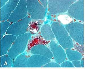
Dilated peri-tubular capillaries filled with sickled RBCs, original Gomori's trichrome stain, × 400. Kanodia et al. Diagnostic Pathology 2008.[1]
The "ragged red fibers" in MELAS syndrome are visible under modified Gomori stain.
It can be used to test for certain forms of mitochondrial myopathy.
It is named for George Gömöri, who developed it in 1950.[4]
References
- Kanodia KV, Vanikar AV, Goplani KR, Gupta SB, Trivedi HL (2008). "Sickle cell nephropathy with diffuse proliferative lupus nephritis: a case report". Diagn Pathol. 3: 9. doi:10.1186/1746-1596-3-9. PMC 2275218. PMID 18307766.
- "GOMORI TRICHROME". Retrieved 2009-04-06.
- "Histology Lab: GOMORI TRICHROME". Retrieved 2009-04-06.
- GOMORI, G. - A rapid one-step trichrome stain. Am. J. Clin. Path. 20: 661-664, 1950.
This article is issued from Wikipedia. The text is licensed under Creative Commons - Attribution - Sharealike. Additional terms may apply for the media files.