Blood culture
A blood culture is a medical laboratory test used to detect bacteria or fungi in a person's blood.[1] Blood is normally sterile, and the presence of microorganisms in the blood often indicates a serious infection (bacteremia, fungemia or sepsis).[2]:863
| Blood culture | |
|---|---|
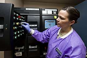
 A medical laboratory technologist unloads blood culture bottles from a Bact-Alert machine, an automated system used to incubate blood cultures and detect bacterial growth | |
| ICD-9 | 90.52 |
| MedlinePlus | 003744 |
The test involves drawing the blood into bottles containing chemicals that encourage microbial growth, which are then placed in an incubator for several days to allow the organisms to multiply.[1] If microbial growth is detected, a Gram stain is made from the blood culture bottle to confirm that bacteria are present and to provide a preliminary identification. The blood is then inoculated onto an agar plate to isolate the organisms for further testing.[3] A positive Gram stain from a blood culture is considered a critical result and must immediately be reported to the clinician.[2]:868–71
To ensure accurate results, blood cultures are drawn using sterile technique. If the sample is contaminated with skin flora, the person will appear to have those organisms in their blood.[2]:868–9 When a blood culture is performed, it is usually drawn in at least two different sets (one set of bottles from each arm) so that contamination is easier to detect. If an organism only appears in one of the two sets, it is more likely to be a contaminant.[1]
Medical uses
When a patient shows signs or symptoms of a systemic infection, results from a blood culture can verify that an infection is present, and they can identify the type (or types) of microorganism that is responsible for the infection. For example, blood tests can identify the causative organisms in severe pneumonia, puerperal fever, pelvic inflammatory disease, neonatal epiglottitis, sepsis, and fever of unknown origin (FUO). However, negative growths do not exclude infection.
Procedure
Collection
Prior to drawing the blood, the tops of the blood culture bottles are disinfected using an alcohol swab,[4] and the venipuncture site is cleaned with alcohol followed by chlorhexidine or 2% tincture of iodine, then left to dry.[note 1] This is done to avoid contamination with organisms from the skin or the environment.[2]:869 A minimum of 10 ml of blood is taken through venipuncture and injected into two or more "blood bottles" with specific media for aerobic and anaerobic organisms. A common medium used for anaerobes is thioglycollate broth. If multiple blood tests need to be drawn at the same time as a blood culture, the blood culture bottles are drawn first to minimize the risk of contamination.[5]:13
To maximise the diagnostic yield of blood cultures, multiple sets of cultures (each set consisting of aerobic and anaerobic vials filled with 3–10 mL) may be obtained. A common protocol used in US hospitals includes the following:
- Set 1 = left antecubital fossa at 0 minutes
- Set 2 = right antecubital fossa at 30 minutes
- Set 3 = left or right antecubital fossa at 90 minutes
Obtaining multiple sets of cultures increases the probability of discovering a pathogenic organism in the blood and reduces the probability of interpreting a positive culture caused by skin contaminants as being due to pathogens.
Culturing

After inoculating the culture vials, advisably with new needles and not the ones used for venipuncture, the vials are sent to the clinical pathology microbiology department. Here the bottles are entered into a blood culture machine, which incubates the specimens at body temperature. The blood culture instrument reports positive blood cultures (cultures with bacteria present, thus indicating the patient is "bacteremic"). Most cultures are monitored for five days, after which negative vials are removed.
If a vial is positive, a microbiologist will perform a Gram stain on the blood for a rapid, general identification of the bacteria, which the microbiologist will report to the attending physician of the bacteremic patient. The blood is also subcultured or "subbed" onto agar plates to isolate the pathogenic organism for culture and susceptibility testing, which takes up to three days. This culture and sensitivity process identifies the species of bacteria. Antibiotic susceptibilities are then assessed on the bacterial isolate to inform clinicians with respect to appropriate antibiotics for treatment.[6] Some guidelines for infective endocarditis recommend taking up to six sets of blood for culture (around 60 ml).
Identification
As of 2011, the pathogens most commonly isolated from blood cultures were coagulase-negative staphylococci, Staphylococcus aureus, Enterococcus species, and Candida albicans.[2]:866
Limitations
Blood cultures are subject to both false positive and false negative errors. False positive results, which occur about 3%+ of the time, can lead to inappropriate treatment.[7]
False negatives may be caused by collecting an insufficient amount of blood or drawing blood cultures after the person has received antibiotics. Additionally, certain organisms are difficult to culture and may not be detected by routine methods.[4] The volume of blood drawn is considered the most important variable in ensuring that pathogens are detected: the more blood that is collected, the more pathogens are recovered.[4] However, if the amount of blood collected far exceeds the recommended volume, bacterial growth may be inhibited by natural inhibitors present in the blood and a relatively inadequate amount of growth medium in the bottle. Over-filling of blood culture bottles may also contribute to iatrogenic anemia.[2]:869
History
Blood cultures were pioneered in the early 20th century.
Notes
- Chlorhexidine is not used in infants under two months old, and iodine-based antiseptics are contraindicated in low-birth-weight infants.[2]:869
References
- American Association for Clinical Chemistry (15 November 2019). "Blood Culture". Lab Tests Online. Retrieved 12 May 2020.
- Connie R. Mahon; Donald C. Lehman; George Manuselis (18 January 2018). Textbook of Diagnostic Microbiology. Elsevier Health Sciences. ISBN 978-0-323-48212-7.
- "Blood culture". MedlinePlus Medical Encyclopedia. 16 September 2019. Retrieved 12 May 2020.
- Garcia, Robert A.; Spitzer, Eric D.; Beaudry, Josephine; Beck, Cindy; Diblasi, Regina; Gilleeny-Blabac, Michelle; Haugaard, Carol; Heuschneider, Stacy; Kranz, Barbara P.; McLean, Karen; Morales, Katherine L.; Owens, Susan; Paciella, Mary E.; Torregrosa, Edwin (2015). "Multidisciplinary team review of best practices for collection and handling of blood cultures to determine effective interventions for increasing the yield of true-positive bacteremias, reducing contamination, and eliminating false-positive central line–associated bloodstream infections". American Journal of Infection Control. 43 (11): 1222–1237. doi:10.1016/j.ajic.2015.06.030. ISSN 0196-6553.
- Kathleen Deska Pagana; Timothy J. Pagana; Theresa N Pagana (19 September 2014). Mosby's Diagnostic and Laboratory Test Reference - E-Book. Elsevier Health Sciences. p. 13. ISBN 978-0-323-22592-2.
- Bouza E, Sousa D, Rodríguez-Créixems M, et al. (2007). "Is blood volume cultured still important for the diagnosis of bloodstream infections?". Journal of Clinical Microbiology. 45 (9): 2765–2769. doi:10.1128/JCM.00140-07. PMC 2045273. PMID 17567782.
- Madeo M, Davies D, Owen L, Wadsworth P, Johnson G, Martin C (2003). "Reduction in the contamination rate of blood cultures collected by medical staff in the accident and emergency department". Clinical Effectiveness in Nursing. 7: 30–32. doi:10.1016/s1361-9004(03)00041-4.