Biomolecular condensate
Biomolecular condensates are a class of non-membrane bound organelles and organelle subdomains. As with other organelles, biomolecular condensates are specialized subunits of the cell. However, unlike many organelles, biomolecular condensate composition is not controlled by a bounding membrane. Instead they can form through a range of different processes, the most well-known of which is phase separation of proteins, RNA and other biopolymers into colloids.
The term 'colloid' was coined by Thomas Graham to describe the behaviour of certain biological macromolecules and inorganic molecules in 1861,[1] while the physics of phase separation was described by Josiah Willard Gibbs in his landmark paper titled On the Equilibrium of Heterogeneous Substances, published in parts between 1875 and 1878.[2]
The concept of intracellular condensation as an organizing principle for the compartmentalization of living cells dates back to the end of the 19th century, beginning with William Bate Hardy and Edmund Beecher Wilson who described the cytoplasm (then called 'protoplasm') as a colloid.[3][4] Around the same time, Thomas Harrison Montgomery Jr. described the morphology of the nucleolus, an organelle within the nucleus, which has subsequently been shown to form through intracellular phase separation.[5] WB Hardy linked formation of biological colloids with phase separation in his study of globulins, stating that: "The globulin is dispersed in the solvent as particles which are the colloid particles and which are so large as to form an internal phase",[6] and further contributed to the basic physical description of oil-water phase separation.[7] Tanaka & Benedek later identified phase-separation behaviour of gamma-crystallin proteins from cataracts in solution,[8][9][10][11] which Benedek referred to as 'protein condensation'.[12]
Advances in microscopy over the 20th century identified many cellular compartments within the cytoplasm or nucleus which were variously referred to as 'puncta', 'dots', 'granules', 'bodies', 'assemblies', 'paraspeckles', 'droplets', or 'aggregates'. The concept of phase separation was subsequently re-borrowed from colloidal chemistry and proposed to underlie both cytoplasmic and nuclear compartmentalization.[13][14]
The newly coined term "biomolecular condensate"[15] refers to biological polymers (as opposed to synthetic polymers) that undergo self assembly via clustering to increase the local concentration of the assembling components, and is analogous to the physical definition of condensation.[16][15] In physics, condensation typically refers to a gas-liquid phase transition. In biology the term 'condensation' is used much more broadly and can also refer to liquid-liquid, liquid-gel, or liquid-solid phase separation to form colloidal suspensions, as well as liquid-to-solid phase transitions such as DNA condensation during prophase of the cell cycle or protein condensation of crystallins in cataracts.[17] With this in mind, the term 'biomolecular condensates' was deliberately introduced to reflect this breadth (see below). Since biomolecular condensation generally involves oligomeric or polymeric interactions between an indefinite number of components, it is generally considered distinct from formation of smaller stochiometric protein complexes with defined numbers of subunits, such as viral capsids or the proteasome - although both are examples of spontaneous self-assembly or self-organisation.
Since 2008, evidence for biomacromolecules undergoing intracellular phase transitions (phase separation) has been observed in many different contexts, both within cells and in reconstituted in vitro experiments.[18][19][20][21][22][23][24][25][26]
Intracellular phase transitions
The term biomolecular condensates was introduced in the context of intracellular assemblies as a convenient and non-exclusionary term to describe non-stoichiometric assemblies of biomolecules.[15] The choice of language here is specific and important. It has been proposed that many biomolecular condensates form through liquid-liquid phase separation (LLPS), liquid-gel phase separation or liquid-solid phase separation;[27] however, unequivocally demonstrating that a cellular body forms through phase separation is challenging.[28][29][30][31] Similarly, while much attention has been paid to the formation of LLPS assemblies, different material states (liquid vs. gel vs. solid) are not always easy to distinguish in living cells.[32][33]
The term "biomolecular condensate" directly address both of these challenges by making no assumption regarding either the physical mechanism through which assembly is achieved, nor the material state of the resulting assembly. Consequently, cellular bodies that form through phase separation are a subset of biomolecular condensates, as are those where the physical origins of assembly are unknown. Historically, many cellular non-membrane bound compartments identified microscopically fall under the broad umbrella of biomolecular condensates.
Further, it appears that intrinsically disordered proteins might play a role in distinguishing between liquid phase states, though how they play this role remains unknown.[27]As of 2019, a light activated system had been developed to experimentally induce condensation.[27]
Examples

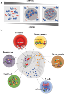
Many examples of biomolecular condensates have been characterized in the cytoplasm and the nucleus.
Cytoplasmic biomolecular condensates thought to arise by either liquid-liquid, liquid-gel or liquid-solid phase separation:
- Lewy bodies
- Stress granule
- P-body
- Germline P-granules
- Lipid droplets
- Starch granules
- Glycogen granules
- Corneal lens formation and cataracts
- Other cytoplasmic inclusions such as pigment granules or cytoplasmic crystals
- Misfolded protein aggregation such as amyloid fibrils or mutant Haemoglobin S (HbS) fibres in sickle cell disease
- Membrane protein, or membrane-associated protein, clustering at neurological synapses, cell-cell junctions, or other membrane domains.
- Other large multiprotein complexes or supramolecular assemblies in the Wnt signaling pathway.[34][35]
It can also be argued that cytoskeletal filaments form by a polymerisation process similar to phase separation, except ordered into filamentous networks instead of amorphous droplets or granules.
Nuclear biomolecular condensates:
Other nuclear structures including heterochromatin and DNA condensation in condensed mitosis chromosomes form by mechanisms similar to phase separation, so can also be classified as biomolecular condensates.
One of the first discovered examples of highly dynamic biomolecular condensate were the supramolecular complexes formed by components of the Wnt signaling pathway.[37][38] The Dishevelled (Dsh) protein undergoes clustering in the cytoplasm via its DIX domain, which mediates protein polymerisation, and is important for signal transduction. The Dsh protein functions both in planar polarity and Wnt signalling, where it recruits another supramolecular complex (the Axin complex) to Wnt receptors at the plasma membrane. The formation of these Dishevelled and Axin containing droplets is conserved across metazoans, including in Drosophila, Xenopus, and human cells.
Another example of liquid droplets in cells are the germline P granules in Caenorhabditis elegans.[27][29] These granules separate out from the cytoplasm and form droplets, as oil does from water. Both the granules and the surrounding cytoplasm are liquid in the sense that they flow in response to forces, and two of the granules can coalesce when they come in contact. When (some of) the molecules in the granules are studied (via fluorescence recovery after photobleaching), they are found to rapidly turnover in the droplets, meaning that molecules diffuse into and out of the granules, just as expected in a liquid droplet. The droplets can also grow to be many molecules across (micrometres)[29] Studies of droplets of the Caenorhabditis elegans protein LAF-1 in vitro[39] also show liquid-like behaviour, with an apparent viscosity Pa s. This is about a ten thousand times that of water at room temperature, but it is small enough to enable the LAF-1 droplets to flow like a liquid.

References
- Graham T (December 1861). "Liquid diffusion applied to analysis". Philosophical Transactions of the Royal Society. 151: 183–224. doi:10.1098/rstl.1861.0011.
- Gibbs, J. W. (1961), Scientific Papers, Dover, New York
- Wilson EB (July 1899). "The Structure of Protoplasm". Science. 10 (237): 33–45. Bibcode:1899Sci....10...33W. doi:10.1126/science.10.237.33. PMID 17829686.
- Hardy WB (May 1899). "On the structure of cell protoplasm: Part I. The Structure produced in a Cell by Fixative and Post-mortem change. The Structure of Colloidal matter and the Mechanism of Setting and of Coagulation". The Journal of Physiology. 24 (2): 158–210.1. doi:10.1113/jphysiol.1899.sp000755. PMC 1516635. PMID 16992486.
- Montgomery T (1898). "Comparative cytological studies, with especial regard to the morphology of the nucleolus". Journal of Morphology. 15 (1): 265–582. doi:10.1002/jmor.1050150204.
- Hardy WB (1905). "Colloidal Solution. The Globulins". Journal of Physiology. 33 (4–5): 255–333. doi:10.1113/jphysiol.1905.sp001126. PMC 1465795. PMID 16992817.
- Hardy WB (1912). "The tension of composite fluid surfaces and the mechanical stability of films of fluid". Proceedings of the Royal Society A. 86 (591): 610–635. doi:10.1098/rspa.1912.0053.
- Tanaka T, Benedek GB (Jun 1975). "Observation of protein diffusivity in intact human and bovine lenses with application to cataract". Investigative Ophthalmology & Visual Science. 14 (6): 449–56. PMID 1132941.
- Tanaka T, Ishimoto C, Chylack LT (Sep 1977). "Phase separation of a protein-water mixture in cold cataract in the young rat lens". Science. 197 (4307): 1010–1012. doi:10.1126/science.887936. PMID 887936.
- Ishimoto C, Goalwin PW, Sun ST, Nishio I, Tanaka T (Sep 1979). "Cytoplasmic phase separation in formation of galactosemic cataract in lenses of young rats". Proceedings of the National Academy of Sciences of the United States of America. 76 (9): 4414–4416. doi:10.1073/pnas.76.9.4414. PMC 411585. PMID 16592709.
- Broide ML, Berland CR, Pande J, Ogun OO, Benedek GB (Jul 1991). "Binary-liquid phase separation of lens protein solutions". Proceedings of the National Academy of Sciences of the United States of America. 88 (13): 5660–4. doi:10.1073/pnas.88.13.5660. PMID 2062844.
- Benedek GB (September 1997). "Cataract as a protein condensation disease: the Proctor Lecture". Investigative Ophthalmology & Visual Science. 38 (10): 1911–21. PMID 9331254.
- Walter H, Brooks DE (March 1995). "Phase separation in cytoplasm, due to macromolecular crowding, is the basis for microcompartmentation". FEBS Letters. 361 (2–3): 135–9. doi:10.1016/0014-5793(95)00159-7. PMID 7698310.
- Iborra FJ (April 2007). "Can visco-elastic phase separation, macromolecular crowding and colloidal physics explain nuclear organisation?". Theoretical Biology & Medical Modelling. 4 (15): 15. doi:10.1186/1742-4682-4-15. PMC 1853075. PMID 17430588.
- Banani SF, Lee HO, Hyman AA, Rosen MK (May 2017). "Biomolecular condensates: organizers of cellular biochemistry". Nature Reviews. Molecular Cell Biology. 18 (5): 285–298. doi:10.1038/nrm.2017.7. PMID 28225081. S2CID 37694361.
- Wheeler RJ, Hyman AA (May 2018). "Controlling compartmentalization by non-membrane-bound organelles". Philosophical Transactions of the Royal Society B: Biological Sciences. 373 (1747): 4666–4684. doi:10.1098/rstb.2017.0193. PMC 5904305. PMID 29632271.
- Benedek GB (September 1997). "Cataract as a protein condensation disease: the Proctor Lecture". Investigative Ophthalmology & Visual Science. 38 (10): 1911–21. PMID 9331254.
- Dumetz AC, Chockla AM, Kaler EW, Lenhoff AM (Jan 2008). "Protein phase behavior in aqueous solutions: crystallization, liquid-liquid phase separation, gels, and aggregates". Biophysical Journal. 94 (2): 570–83. doi:10.1529/biophysj.107.116152. PMC 2157236. PMID 18160663.
- Brangwynne CP, Eckmann CR, Courson DS, Rybarska A, Hoege C, Gharakhani J, et al. (June 2009). "Germline P granules are liquid droplets that localize by controlled dissolution/condensation". Science. 324 (5935): 1729–32. Bibcode:2009Sci...324.1729B. doi:10.1126/science.1172046. PMID 19460965.
- Larson AG, Elnatan D, Keenen MM, Trnka MJ, Johnston JB, Burlingame AL, et al. (July 2017). "Liquid droplet formation by HP1α suggests a role for phase separation in heterochromatin". Nature. 547 (7662): 236–240. Bibcode:2017Natur.547..236L. doi:10.1038/nature22822. PMC 5606208. PMID 28636604.
- Nott TJ, Petsalaki E, Farber P, Jervis D, Fussner E, Plochowietz A, et al. (March 2015). "Phase transition of a disordered nuage protein generates environmentally responsive membraneless organelles". Molecular Cell. 57 (5): 936–947. doi:10.1016/j.molcel.2015.01.013. PMC 4352761. PMID 25747659.
- Patel A, Lee HO, Jawerth L, Maharana S, Jahnel M, Hein MY, et al. (August 2015). "A Liquid-to-Solid Phase Transition of the ALS Protein FUS Accelerated by Disease Mutation". Cell. 162 (5): 1066–77. doi:10.1016/j.cell.2015.07.047. PMID 26317470.
- Feric M, Vaidya N, Harmon TS, Mitrea DM, Zhu L, Richardson TM, et al. (June 2016). "Coexisting Liquid Phases Underlie Nucleolar Subcompartments". Cell. 165 (7): 1686–1697. doi:10.1016/j.cell.2016.04.047. PMC 5127388. PMID 27212236.
- Riback JA, Zhu L, Ferrolino MC, Tolbert M, Mitrea DM, Sanders DW, et al. (2019-10-22). "Composition dependent phase separation underlies directional flux through the nucleolus". bioRxiv: 809210. doi:10.1101/809210.
- Li P, Banjade S, Cheng HC, Kim S, Chen B, Guo L, et al. (March 2012). "Phase transitions in the assembly of multivalent signalling proteins". Nature. 483 (7389): 336–40. Bibcode:2012Natur.483..336L. doi:10.1038/nature10879. PMC 3343696. PMID 22398450.
- Feric M, Vaidya N, Harmon TS, Mitrea DM, Zhu L, Richardson TM, et al. (June 2016). "Coexisting Liquid Phases Underlie Nucleolar Subcompartments". Cell. 165 (7): 1686–1697. doi:10.1016/j.cell.2016.04.047. PMC 5127388. PMID 27212236.
- Tang, Lei (February 2019). "Optogenetic tools light up phase separation". Nature Methods (Paper). 16 (2): 139. doi:10.1038/s41592-019-0310-5. PMID 30700901.(subscription required)
- Hyman AA, Weber CA, Jülicher F (2014-10-11). "Liquid-liquid phase separation in biology". Annual Review of Cell and Developmental Biology. 30 (1): 39–58. doi:10.1146/annurev-cellbio-100913-013325. PMID 25288112.
- Brangwynne CP, Eckmann CR, Courson DS, Rybarska A, Hoege C, Gharakhani J, et al. (June 2009). "Germline P granules are liquid droplets that localize by controlled dissolution/condensation". Science. 324 (5935): 1729–32. Bibcode:2009Sci...324.1729B. doi:10.1126/science.1172046. PMID 19460965.
- McSwiggen DT, Mir M, Darzacq X, Tjian R (December 2019). "Evaluating phase separation in live cells: diagnosis, caveats, and functional consequences". Genes & Development. 33 (23–24): 1619–1634. doi:10.1101/gad.331520.119. PMC 6942051. PMID 31594803.
- Posey AE, Holehouse AS, Pappu RV (2018). "Phase Separation of Intrinsically Disordered Proteins". Methods in Enzymology. Elsevier. 611: 1–30. doi:10.1016/bs.mie.2018.09.035. ISBN 978-0-12-815649-0. PMID 30471685.
- Woodruff JB, Hyman AA, Boke E (February 2018). "Organization and Function of Non-dynamic Biomolecular Condensates". Trends in Biochemical Sciences. 43 (2): 81–94. doi:10.1016/j.tibs.2017.11.005. PMID 29258725.
- Boeynaems S, Alberti S, Fawzi NL, Mittag T, Polymenidou M, Rousseau F, et al. (June 2018). "Protein Phase Separation: A New Phase in Cell Biology". Trends in Cell Biology. 28 (6): 420–435. doi:10.1016/j.tcb.2018.02.004. PMC 6034118. PMID 29602697.
- Schaefer KN, Peifer M (February 2019). "Wnt/Beta-Catenin Signaling Regulation and a Role for Biomolecular Condensates". Developmental Cell. 48 (4): 429–444. doi:10.1016/j.devcel.2019.01.025. PMC 6386181. PMID 30782412.
- Gammons M, Bienz M (April 2018). "Multiprotein complexes governing Wnt signal transduction". Current Opinion in Cell Biology. 51 (1): 42–49. doi:10.1016/j.ceb.2017.10.008. PMID 29153704.
- Feric M, Vaidya N, Harmon TS, Mitrea DM, Zhu L, Richardson TM, et al. (June 2016). "Coexisting Liquid Phases Underlie Nucleolar Subcompartments". Cell. 165 (7): 1686–1697. doi:10.1016/j.cell.2016.04.047. PMC 5127388. PMID 27212236.
- Schaefer KN, Peifer M (February 2019). "Wnt/Beta-Catenin Signaling Regulation and a Role for Biomolecular Condensates". Developmental Cell. 48 (4): 429–444. doi:10.1016/j.devcel.2019.01.025. PMC 6386181. PMID 30782412.
- Gammons M, Bienz M (April 2018). "Multiprotein complexes governing Wnt signal transduction". Current Opinion in Cell Biology. 51 (1): 42–49. doi:10.1016/j.ceb.2017.10.008. PMID 29153704.
- Elbaum-Garfinkle S, Kim Y, Szczepaniak K, Chen CC, Eckmann CR, Myong S, Brangwynne CP (June 2015). "The disordered P granule protein LAF-1 drives phase separation into droplets with tunable viscosity and dynamics". Proceedings of the National Academy of Sciences of the United States of America. 112 (23): 7189–94. Bibcode:2015PNAS..112.7189E. doi:10.1073/pnas.1504822112. PMC 4466716. PMID 26015579.
Further reading
- Ditlev JA, Case LB, Rosen MK (November 2018). "Who's In and Who's Out-Compositional Control of Biomolecular Condensates". Journal of Molecular Biology. 430 (23): 4666–4684. doi:10.1016/j.jmb.2018.08.003. PMC 6204295. PMID 30099028.
- Banani SF, Lee HO, Hyman AA, Rosen MK (May 2017). "Biomolecular condensates: organizers of cellular biochemistry". Nature Reviews. Molecular Cell Biology. 18 (5): 285–298. doi:10.1038/nrm.2017.7. PMID 28225081. S2CID 37694361.
- Hyman AA, Weber CA, Jülicher F (2014). "Liquid-liquid phase separation in biology". Annual Review of Cell and Developmental Biology. 30: 39–58. doi:10.1146/annurev-cellbio-100913-013325. PMID 25288112.
- Dolgin E (March 2018). "What lava lamps and vinaigrette can teach us about cell biology". Nature. 555 (7696): 300–302. Bibcode:2018Natur.555..300D. doi:10.1038/d41586-018-03070-2. PMID 29542707.