Vibratome
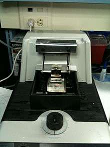
A vibratome is an instrument used to cut thin slices of material. It is similar to a microtome but uses a vibrating blade to cut through tissue. The vibration amplitude, the speed, and the angle of the blade can all be controlled. Fixed or fresh tissue pieces are embedded in low gelling temperature agarose.(Some have had success without using the agarose to embed) The resulting agarose block containing the tissue piece is then attached to a metal block and sectioned while submerged in a water or buffer bath. Individual sections are then collected with a fine brush and transferred to slides or multiwell plates for staining.[1]
Types of vibratomes
Currently, there are multiple types of vibrating microtomes for use by laboratories and industry labs around the world. The following is a list of the most commonly used vibratomes, and their advantages and disadvantages.
Compresstome vibratome
Compresstome vibratomes are designed and made by Precisionary Instruments,[2] which is headquartered in Boston, MA, and were introduced to the market in 2004. The vibratome models include the VF-200, VF-200-0Z, VF-300, VF-300-0Z, and VF-700-0Z.[3] The "-0Z" term on the models refer to the "Auto Zero-Z" patent pending technology[4] developed by Precisionary, where these models are calibrated during the manufacturing process to have near-zero deflections or vibrations in the z-axis. Auto Zero-Z therefore allows tissues to be cut without z-axis shearing of the specimen surface, which helps to yield healthier cells. For larger specimens, such as sectioning tissues from sheep, bovine, pigs, and non-human primates, the company also offers vibratome models VF-800-0Z and VF-900-0Z.
Applications of the Compresstome vibratomes include making slices for electrophysiology, immunohistochemistry, precision-cut slices (lung, liver, intestines, heart, kidney, etc), mature animal tissues, and organotypic cultures.[5] Organ systems that use Compresstome vibratomes for research include adipose (fat), heart,[6] brain, liver, lung, kidney, muscle, lymph nodes, and gastrointestinal tissues. The Compresstome can section tissue slices with a thickness range of 3 μm to 1000 μm, and therefore is capable of sectioning tissue as thin as cryostats but without the freeze-thaw cycle of tissue preparation.
The Compresstome patented technology works by embedding tissue (both fixed and live specimens) in agarose inside a specimen tube. The tube has one end that is narrower than the other, so that when the embedded tissue is gently pushed out for cutting, there is a slight "compression" of the tissue. The compression puts a slight amount of pressure on the tissue and agarose, which helps to hold the specimen in place and prevents the vibrating knife from pushing against the tissue. As a result, this patented technology prevents tissue shearing or tearing, and thus prevents the introduction of "chattermarks" or vibration artifacts during the cutting process.[7]
Compresstome vibratomes function similarly to Leica vibratomes, but are in the low- to mid-cost category of laboratory equipment, making them more affordable for research labs. Compresstome machines weight 5–9 kg, meaning that they have a small footprint and are easily portable and moveable.
Leica vibratome
Leica vibratomes include the models VT1000S, VT1200, and VT1200S.[8] In 2008, Leica Microsystems acquired the company McCormick Scientific (a St. Louis, Missouri-based company also called Coretech Holdings) which owned The Vibratome Company.[9] Today, the original Vibratome is no longer produced, and labs with the Vibratome are no longer able to get it serviced by Leica. The newer models are available and are vibrating blade microtomes used for sectioning fixed and live tissues. Applications include neurophysiology, neuropathology, and experimental pathology. Tissue section thickness ranges from 100 μm to 300 μm for live tissue, although if the section thickness chosen is too thin, no cells remain intact. Thicknesses down to 10-20 μm can be achieved with fixed tissues. Thicknesses of 2-3 μm that are achieved with a rotary microtome or cryostat cannot be achieved with a vibrating blade microtome.[10]
Although tissue embedding is not necessary, it is highly recommended that very small and soft specimens be embedded in agar or agarose.[10] Embedding the tissue has no effect on the tissue itself, and agar or agarose embedding can be used for both live and fixed tissues. This process is very similar to the Compresstome vibratome procedures. One commonly encountered problem with the Leica vibratome is that the cutting knife pushes away the specimen which results in shearing or tearing of tissue. An agar block can be glued below the tissue specimen in an attempt to overcome this obstacle.
When the vibrating knife pushes against the tissue, it can lead to the most common problem for the Leica slicers: Scientists frequently report getting "thick and thin" sections, where the first section is really thin or nothing, and the next slice is very thick or double what it should be. The alternating "thick and thin" slices are a challenge because electrophysiology and immunohistochemistry experiments require evenly cut slices with consistent thicknesses for reproducible studies. The cause of the "thick and thin" problem is attributed to technique and high knife cutting speed. Decreasing the cutting speed may help resolve this problem, but also results in slower cutting times.[10] Thus, it is not uncommon for a researcher to wait several minutes for each cut section.
Leica vibratomes are the most expensive vibrating microtomes available, placing them in the high-cost category of laboratory instruments. Vibratome machines also weigh about 20–25 kg, making them heavy to move so a designated area of sectioning in the lab is required.
Campden Instruments vibratome
Sectioning with Campden vibratomes have been reported for over 30 years. The most recent models include the 7000smz-2, the 5100mz, and the 5100mz-Plus. All three models have operating modes that are manual and semi-automated for the slice window.[11] The previous slicer models were called Integraslice, but these machines have since been discontinued. The 7000smz-2 model is equivalent to the Compresstome VF-300-0Z and the Leica 1200S. Keeping tissues cool during the cutting process can be done with passive cooling such as using ice or buffer solution slush. Campden vibratomes weight about 15 kg and cost more than Compresstome vibratomes but less than Leica microtomes.
Advantages
- No need to dehydrate tissues prior to embedding, thus decreased loss of cell constituents
- No messy paraffin embedding
- Allows for agarose embedding of tissue to provide cutting stability
- Compresstome microtomes are a subtype of vibratomes that using gentle agarose-embedded compression technology to prevent chattermarks, shearing, and tearing of tissue during the cutting process[12]
- Compresstome vibratomes overcome artifacts such as ice crystals that form during cryostat embedding procedures
- No need to deparaffinise and rehydrate sections prior to immunostaining
- No high temperatures or harsh chemical treatments that may lead to antigen instability
- No special microtome blades required
- Compresstome vibratomes better preserve cellular architecture from older or more mature animals, where myelination makes tissue more difficult to cut[1]
- Less chance of artifacts caused by parrafin embedding or freezing
- Decreased tissue autofluoresecence due to avoidance of formalin-fixation and paraffin embedding
- Less wait period from tissue sampling to time of immunolabelling
- Allows for direct creation of free-floating sections for immunohistochemistry
Disadvantages
- Instead of ribbons, single sections are cut and collected which are more delicate and difficult to handle.
- Sections are generally thicker than those obtained with paraffin methods; penetration of antibodies and other reagents may be slower and thus longer incubation times may be necessary. Also, thick sections may be difficult to image with the microscope. (However, thick sections are compatible and sometimes even desirable if using confocal microscopy.)
- Leica vibratomes are expensive and heavy, preventing simple mobility of the machine in a laboratory setting.[12]
- Securing vibratome sections to glass slides can be difficult or impossible, due to the thickness of the sections.[13]
References
- 1 2 "Improved methods for acute brain slice preparation from adult and aging animals" (PDF). Archived from the original (PDF) on July 17, 2011. Retrieved June 1, 2009.
- ↑ "Precisionary Instruments Company History". www.precisionary.com. Retrieved 2016-09-07.
- ↑ "Compresstome models".
- ↑ "Auto Zero-Z Technology".
- ↑ "Multiple applications of Compresstome for research".
- ↑ "Sectioning live myocardial slices with Compresstome VF-300 For Tissue Culture".
- ↑ Abdelaal, Hadia M; Kim, Hyeon O; Wagstaff, Reece; Sawahata, Ryoko; Southern, Peter J; Skinner, Pamela J (2015). "Comparison of Vibratome and Compresstome sectioning of fresh primate lymphoid and genital tissues for in situ MHC-tetramer and immunofluorescence staining". Biological Procedures Online. 17 (1): 2. doi:10.1186/s12575-014-0012-4. PMC 4318225. PMID 25657614.
- ↑ "Vibrating Blade Microtome: Leica Biosystems". Leica Biosystems. Retrieved 2016-09-07.
- ↑ "Leica Microsystems acquires Coretech Holdings/McCormick Scientific". Retrieved 2016-09-07.
- 1 2 3 "Leica VT1000 S - FAQ: Leica Biosystems". Leica Biosystems. Retrieved 2016-09-07.
- ↑ "Campden Vibrating Microtomes | Campden Instruments LTD". www.campdeninstruments.com. Retrieved 2016-09-07.
- 1 2 "Can anyone provide advice on purchasing a vibratome for..." www.researchgate.net. Retrieved 2016-09-06.
- ↑ http://www4.ncsu.edu/~rgfranks/FRANKS%20LAB%20PROTOCOLS/immuno%20histio%20chemistry/robertson%20vbratmimuno.doc%5Bpermanent+dead+link%5D