TUBA1A


Tubulin alpha-1A chain is a protein that in humans is encoded by the TUBA1A gene.[5][6][7]
Background
TUBA1A is a structural gene that encodes for Tubulin, Alpha 1A product. TUBA1A product is an alpha-tubulin that participates in the formation of microtubules - structural proteins that participate in cytoskeletal structure. Specifically, microtubules are composed of a heterodimer of alpha and beta-tubulin molecules. Cowan et al. demonstrated that bα1 is a primary α-tubulin of the human fetal brain, and that it is expressed solely in that structure, by way of Northern blot.[8] Miller et al. further elaborated on the role of α-tubulins and the process of neuronal development and maturation, comparing the expressions of rat α-tubulins Tα1 and T26. These two rat α-tubulins are homologs of bα1 and kα1 showing that a rat homolog of human TUBA1A (Tα1) had elevated expression during the extension of neuronal processes. Culturing of pheochromocytoma cells with Nerve Growth Factor (NGF) induced differentiation and the development of neuronal processes. Northern blot assay showed markedly elevated levels of Tα1 mRNA expression; T26 mRNA expression increased minimally with exposure to NGF.[9] These data suggest that TUBA1A models the brain by participating in the directing of neuronal migration through the ability of microtubules to readily form and break polymers to extend and retract processes to induce nucleokinesis.[10] Poirier et al. used RNA in situ hybridization to show TUBA1A expression in mice embryo; embryo sections from embryonic day 16.5 “showed a strong labeling in the telencephalon, diencephalon, and mesencephalon, the developing cerebellum, the brainstem, the spinal cord, and the dorsal root ganglia”.[11]
Function
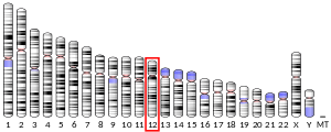
Microtubules of the eukaryotic cytoskeleton perform essential and diverse functions and are composed of a heterodimer of alpha and beta tubulins. The genes encoding these microtubule constituents belong to the tubulin superfamily, which is composed of six distinct families. Genes from the alpha, beta and gamma tubulin families are found in all eukaryotes. The alpha and beta tubulins represent the major components of microtubules, while gamma tubulin plays a critical role in the nucleation of microtubule assembly. There are multiple alpha and beta tubulin genes, which are highly conserved among species. This gene encodes alpha tubulin and is highly similar to mouse and rat Tuba1 gene. Northern blotting studies have shown that the gene expression is predominantly found in morphologically differentiated neurologic cells. This gene is one of three alpha-tubulin genes in a cluster on chromosome 12q.[7]
Interactions
Disease
Mutations to the TUBA1A gene manifest clinically as Type 3 Lissencephaly. In general, lissencephaly is characterized by agyria (lacking of gyri and sulci to the brain – a smooth brain), seizure activity, failure to thrive, as well as intellectual disability and psychomotor retardation, often to a profound degree.[11] The symptoms of Lis3 Lissencephaly are not especially different from generalized lissencephaly (Lis1, related to PAFAH1B1). Diagnosis of lissencephaly generally is made from the symptom profile, while attribution to a specific type is obtained by microarray. Treatment is symptomatic; anti-convulsive drugs for seizure activity, g-button gastrostomy to feed the child, physical therapy for muscle disorders. TUBA1A mutation is common in microlissencephaly
Animal Model
Keays et al. describe a mouse with a mutation of the TUBA1A gene induced by N-ethyl-N-nitrosourea. The relevant point mutation resulted in S140G;[13] the site of the mutation participates in the N-site of the formed α-tubulin, and participates in stabilizing the α-β tubulin polymer by binding GTP at this site.[14] The S140G mutation resulted in the formation of a “compromised GTP binding pocket”. Authors note defects associated with cortical layers II/III and IV, especially in cortical neuronal migration (with respect to wild-type counterparts), showing that the S140G mutation has value as a model for detailing disease associated with the Human TUBA homolog.[13]
References
- 1 2 3 GRCh38: Ensembl release 89: ENSG00000167552 - Ensembl, May 2017
- 1 2 3 GRCm38: Ensembl release 89: ENSMUSG00000072235 - Ensembl, May 2017
- ↑ "Human PubMed Reference:".
- ↑ "Mouse PubMed Reference:".
- ↑ Crabtree DV, Ojima I, Geng X, Adler AJ (August 2001). "Tubulins in the primate retina: evidence that xanthophylls may be endogenous ligands for the paclitaxel-binding site". Bioorganic & Medicinal Chemistry. 9 (8): 1967–76. doi:10.1016/S0968-0896(01)00103-1. PMID 11504633.
- ↑ Hall JL, Cowan NJ (January 1985). "Structural features and restricted expression of a human alpha-tubulin gene". Nucleic Acids Research. 13 (1): 207–23. doi:10.1093/nar/13.1.207. PMC 340985. PMID 3839072.
- 1 2 "Entrez Gene: TUBA1A tubulin, alpha 1a".
- ↑ Cowan, N. J.; Dobner, P. R.; Fuchs, E. V.; Cleveland, D. W. (1983). "Expression of Human α-Tubulin Genes: Interspecies Conversion of 3' Untranslated Regions". Molecular and Cell Biology. 3 (10): 1738–1739, 1742. doi:10.1128/mcb.3.10.1738. PMC 370035. PMID 6646120.
- ↑ Mill, F. D.; Naus, C. C.; Durand, M.; Bloom, F. E.; Milner, R. J. (1987). "Isotypes of alpha-tubulin are differentially regulated during neuronal maturation". The Journal of Cell Biology. 105 (6): 3065–3073. doi:10.1083/jcb.105.6.3065. PMC 2114727. PMID 3693406.
- ↑ Sakakaibara, A.; Ando, R.; Spair, T.; Tanaka, T. (July 2013). "Microtubule dynamics in neuronal morphogenesis". Open Biology. 3 (7): 1–2. doi:10.1098/rsob.130061. PMC 3728923. PMID 23864552.
- 1 2 Poirier, K.; Keays, D. A.; Francis, F.; Saillour, Y.; Bahi, N.; Manouvrier, S.; Fallet-Bianco, C.; Paquier, L.; Toutain, A.; Tuy, F. P. D.; Bienvenu, T.; Joriot, S.; Odent, S.; Ville, D.; Desguerre, I.; Goldenberg, A.; Moutard, M.-L.; Fryns, J.-P.; van Esch, H.; Harvey, R. J.; Siebold, C.; Flint, J.; Beldjord, C.; Chelly, J. (November 2007). "Large Spectrum of Lissencephaly and Pachygyria Phenotypes Resulting from De Novo Missense Mutations in Tubulin Alpha 1A (TUBA1A)". Human Mutation. 28 (11): 1058–1061. doi:10.1002/humu.20572. PMID 17584854.
- ↑ Sapir T, Elbaum M, Reiner O (December 1997). "Reduction of microtubule catastrophe events by LIS1, platelet-activating factor acetylhydrolase subunit". The EMBO Journal. 16 (23): 6977–84. doi:10.1093/emboj/16.23.6977. PMC 1170301. PMID 9384577.
- 1 2 Keays, D. A.; Tian, G.; Poirier, K.; Huang, G.-J.; Siebold, C.; Cleak, J.; Oliver, P. L.; Fray, M.; Harvey, R. J.; Molnár, Z.; Piñon, M. C.; Dear, N.; Valdar, W.; Brown, S. D.; Davies, K. E.; Rawlins, J. N. P.; Cowan, N. J.; Nolan, P.; Chelly, J.; Flint, J. (January 2007). "Mutations in α-Tubulin Cause Abnormal Neuronal Migration in Mice and Lissencephaly in Humans". Cell. 128 (1): 45–46, 48–50. doi:10.1016/j.cell.2006.12.017. PMC 1885944. PMID 17218254.
- ↑ Löwe, J.; Li, H.; Downing, K. H.; Nogales, E. (November 2001). "Refined structure of αβ-tubulin at 3.5 Å resolution". Journal of Molecular Biology. 313 (5): 1045–1046. doi:10.1006/jmbi.2001.5077. PMID 11700061.
Further reading
- Desai A, Mitchison TJ (July 1998). "Tubulin and FtsZ structures: functional and therapeutic implications". BioEssays. 20 (7): 523–7. doi:10.1002/(SICI)1521-1878(199807)20:7<523::AID-BIES1>3.0.CO;2-L. PMID 9722999.
- Oakley BR (December 2000). "An abundance of tubulins". Trends in Cell Biology. 10 (12): 537–42. doi:10.1016/S0962-8924(00)01857-2. PMID 11121746.
- Dutcher SK (February 2001). "The tubulin fraternity: alpha to eta". Current Opinion in Cell Biology. 13 (1): 49–54. doi:10.1016/S0955-0674(00)00173-3. PMID 11163133.
- Miller FD, Naus CC, Durand M, Bloom FE, Milner RJ (December 1987). "Isotypes of alpha-tubulin are differentially regulated during neuronal maturation". The Journal of Cell Biology. 105 (6 Pt 2): 3065–73. doi:10.1083/jcb.105.6.3065. PMC 2114727. PMID 3693406.
- Cowan NJ, Dobner PR, Fuchs EV, Cleveland DW (October 1983). "Expression of human alpha-tubulin genes: interspecies conservation of 3' untranslated regions". Molecular and Cellular Biology. 3 (10): 1738–45. doi:10.1128/mcb.3.10.1738. PMC 370035. PMID 6646120.
- Alexandrova N, Niklinski J, Bliskovsky V, Otterson GA, Blake M, Kaye FJ, Zajac-Kaye M (September 1995). "The N-terminal domain of c-Myc associates with alpha-tubulin and microtubules in vivo and in vitro". Molecular and Cellular Biology. 15 (9): 5188–95. PMC 230766. PMID 7651436.
- Sapir T, Elbaum M, Reiner O (December 1997). "Reduction of microtubule catastrophe events by LIS1, platelet-activating factor acetylhydrolase subunit". The EMBO Journal. 16 (23): 6977–84. doi:10.1093/emboj/16.23.6977. PMC 1170301. PMID 9384577.
- Kinnunen T, Kaksonen M, Saarinen J, Kalkkinen N, Peng HB, Rauvala H (April 1998). "Cortactin-Src kinase signaling pathway is involved in N-syndecan-dependent neurite outgrowth". The Journal of Biological Chemistry. 273 (17): 10702–8. doi:10.1074/jbc.273.17.10702. PMID 9553134.
- Faruki S, Geahlen RL, Asai DJ (July 2000). "Syk-dependent phosphorylation of microtubules in activated B-lymphocytes". Journal of Cell Science. 113 (14): 2557–65. PMID 10862713.
- Watts NR, Sackett DL, Ward RD, Miller MW, Wingfield PT, Stahl SS, Steven AC (July 2000). "HIV-1 rev depolymerizes microtubules to form stable bilayered rings". The Journal of Cell Biology. 150 (2): 349–60. doi:10.1083/jcb.150.2.349. PMC 2180222. PMID 10908577.
- Germani A, Bruzzoni-Giovanelli H, Fellous A, Gisselbrecht S, Varin-Blank N, Calvo F (December 2000). "SIAH-1 interacts with alpha-tubulin and degrades the kinesin Kid by the proteasome pathway during mitosis". Oncogene. 19 (52): 5997–6006. doi:10.1038/sj.onc.1204002. PMID 11146551.
- Payton JE, Perrin RJ, Clayton DF, George JM (November 2001). "Protein-protein interactions of alpha-synuclein in brain homogenates and transfected cells". Brain Research. Molecular Brain Research. 95 (1–2): 138–45. doi:10.1016/S0169-328X(01)00257-1. PMID 11687285.
- Bifulco M, Laezza C, Stingo S, Wolff J (February 2002). "2',3'-Cyclic nucleotide 3'-phosphodiesterase: a membrane-bound, microtubule-associated protein and membrane anchor for tubulin". Proceedings of the National Academy of Sciences of the United States of America. 99 (4): 1807–12. doi:10.1073/pnas.042678799. PMC 122275. PMID 11842207.
- Saugstad JA, Yang S, Pohl J, Hall RA, Conn PJ (March 2002). "Interaction between metabotropic glutamate receptor 7 and alpha tubulin". Journal of Neurochemistry. 80 (6): 980–8. doi:10.1046/j.0022-3042.2002.00778.x. PMC 2925652. PMID 11953448.
- Ivings L, Pennington SR, Jenkins R, Weiss JL, Burgoyne RD (May 2002). "Identification of Ca2+-dependent binding partners for the neuronal calcium sensor protein neurocalcin delta: interaction with actin, clathrin and tubulin". The Biochemical Journal. 363 (Pt 3): 599–608. doi:10.1042/0264-6021:3630599. PMC 1222513. PMID 11964161.
PDB gallery | |
|---|---|
 1ffx: TUBULIN:STATHMIN-LIKE DOMAIN COMPLEX  1ia0: KIF1A HEAD-MICROTUBULE COMPLEX STRUCTURE IN ATP-FORM 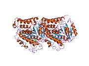 1jff: Refined structure of alpha-beta tubulin from zinc-induced sheets stabilized with taxol 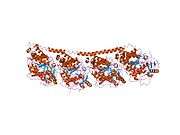 1sa0: TUBULIN-COLCHICINE: STATHMIN-LIKE DOMAIN COMPLEX 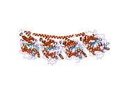 1sa1: Tubulin-podophyllotoxin: stathmin-like domain complex 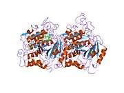 1tub: TUBULIN ALPHA-BETA DIMER, ELECTRON DIFFRACTION 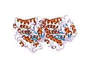 1tvk: The binding mode of epothilone A on a,b-tubulin by electron crystallography 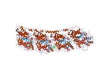 1z2b: Tubulin-colchicine-vinblastine: stathmin-like domain complex 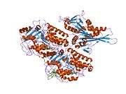 2hxf: KIF1A head-microtubule complex structure in amppnp-form  2hxh: KIF1A head-microtubule complex structure in adp-form |