Prosaposin


Prosaposin, also known as PSAP, is a protein which in humans is encoded by the PSAP gene.[5]
This highly conserved glycoprotein is a precursor for 4 cleavage products: saposins A, B, C, and D. Saposin is an acronym for Sphingolipid Activator PrO[S]teINs.[6] Each domain of the precursor protein is approximately 80 amino acid residues long with nearly identical placement of cysteine residues and glycosylation sites. Saposins A-D localize primarily to the lysosomal compartment where they facilitate the catabolism of glycosphingolipids with short oligosaccharide groups. The precursor protein exists both as a secretory protein and as an integral membrane protein and has neurotrophic activities.[5]
Saposins A-D are required for the hydrolysis of certain sphingolipids by specific lysosomal hydrolases.[7]
Family members
- Saposin A was identified as an N-terminal domain in the prosaposin cDNA prior to its isolation. It is known to stimulate the enzymatic hydrolysis of 4-methlyumbelliferyl-β-glucoside, glucocerebroside, and galactocerebroside.[8]
- Saposin B was the first to be discovered and was found to be required as a heat-stable factor for hydrolysis of sulfatides by arylsulfatase A. It is known by many different names, such as, sphingolipid activator protein-1 (SAP-1), sulfatide activator protein, GM1 ganglioside activator, dispersin, and nonspecific.[9] It has been observed that this particular saposin activates many enzymes through interaction with the substrates not the enzymes themselves.
- Saposin C was the second saposin to be discovered and stimulates the hydrolysis of glycocerebroside by glycosylceramidase and galactocerebroside by galactosylceramidase.
- Saposin D is not well known to due lack of investigation at this point in time. It was predicted from the cDNA sequence of prosaposin, like saposin A. Enzymatic stimulation is very specific for this particular glycoprotein and it not understood completely.[7]
- GM2A (GM2 ganglioside activator) has been viewed as a member of the SAP family and has been called SAP-3 (sphingolipid activator protein 3)[10]
Structure
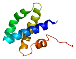
Every saposin contains about 80 amino acid residues and has six equally placed cysteines, two prolines, and a glycosylation site (two in saposin A, one each in saposins B, C, and D).[7] Since saposins characteristics of extreme heat-stability, adundance of disulfide linkages, and resistance to most proteases, they are assumed to be extremely compact and rigidly disulfide-linked molecules. Each saposin has an α-helical structure that is seen as being important for stimulation because this structure is maximal at a pH of 4.5; which is optimal for many lysosomal hydrolases.[7] This helical structure is seen in all (especially with the first region), but saposin has been predicted to have β-sheet configuration due to it first 24 amino acids of the N-end.[9]
Function
They probably act by isolating the lipid substrate from the membrane surroundings, thus making it more accessible to the soluble degradative enzymes. which contains four Saposin-B domains, yielding the active saposins after proteolytic cleavage, and two Saposin-A domains that are removed in the activation reaction. The Saposin-B domains also occur in other proteins, many of them active in the lysis of membranes.[14][15]
Clinical significance
Mutations in this gene have been associated with Gaucher disease, Tay-Sachs disease, and metachromatic leukodystrophy.[6]
See also
References
- 1 2 3 GRCh38: Ensembl release 89: ENSG00000197746 - Ensembl, May 2017
- 1 2 3 GRCm38: Ensembl release 89: ENSMUSG00000004207 - Ensembl, May 2017
- ↑ "Human PubMed Reference:".
- ↑ "Mouse PubMed Reference:".
- 1 2 "Entrez Gene: PSAP prosaposin (variant Gaucher disease and variant metachromatic leukodystrophy)".
- 1 2 Morimoto S, Yamamoto Y, O'Brien JS, Kishimoto Y (May 1990). "Distribution of saposin proteins (sphingolipid activator proteins) in lysosomal storage and other diseases". Proc. Natl. Acad. Sci. U.S.A. 87 (9): 3493–7. doi:10.1073/pnas.87.9.3493. PMC 53927. PMID 2110365.
- 1 2 3 4 Kishimoto Y, Hiraiwa M, O'Brien JS (September 1992). "Saposins: structure, function, distribution, and molecular genetics". J. Lipid Res. 33 (9): 1255–67. PMID 1402395.
- ↑ Morimoto S, Martin BM, Yamamoto Y, Kretz KA, O'Brien JS, Kishimoto Y (May 1989). "Saposin A: second cerebrosidase activator protein". Proc. Natl. Acad. Sci. U.S.A. 86 (9): 3389–93. doi:10.1073/pnas.86.9.3389. PMC 287138. PMID 2717620.
- 1 2 O'Brien JS, Kishimoto Y (March 1991). "Saposin proteins: structure, function, and role in human lysosomal storage disorders". FASEB J. 5 (3): 301–8. PMID 2001789.
- ↑ HUGO Gene Nomenclature Committee, "GM2A", HGNC database, retrieved 2016-03-13.
- ↑ Ahn VE, Leyko P, Alattia JR, Chen L, Privé GG (August 2006). "Crystal structures of saposins A and C". Protein Sci. 15 (8): 1849–57. doi:10.1110/ps.062256606. PMC 2242594. PMID 16823039.
- ↑ Ahn VE, Faull KF, Whitelegge JP, Fluharty AL, Privé GG (January 2003). "Crystal structure of saposin B reveals a dimeric shell for lipid binding". Proc. Natl. Acad. Sci. U.S.A. 100 (1): 38–43. doi:10.1073/pnas.0136947100. PMC 140876. PMID 12518053.
- 1 2 Rossmann M, Schultz-Heienbrok R, Behlke J, Remmel N, Alings C, Sandhoff K, Saenger W, Maier T (May 2008). "Crystal structures of human saposins C and D: implications for lipid recognition and membrane interactions". Structure. 16 (5): 809–17. doi:10.1016/j.str.2008.02.016. PMID 18462685.
- ↑ Ponting CP (1994). "Acid sphingomyelinase possesses a domain homologous to its activator proteins: saposins B and D". Protein Sci. 3 (2): 359–361. doi:10.1002/pro.5560030219. PMC 2142785. PMID 8003971.
- ↑ Hofmann K, Tschopp J (1996). "Cytotoxic T cells: more weapons for new targets?". Trends Microbiol. 4 (3): 91–94. doi:10.1016/0966-842X(96)81522-8. PMID 8868085.
Further reading
- Gieselmann V, Zlotogora J, Harris A, et al. (1995). "Molecular genetics of metachromatic leukodystrophy". Hum. Mutat. 4 (4): 233–42. doi:10.1002/humu.1380040402. PMID 7866401.
- Schnabel D, Schröder M, Fürst W, et al. (1992). "Simultaneous deficiency of sphingolipid activator proteins 1 and 2 is caused by a mutation in the initiation codon of their common gene". J. Biol. Chem. 267 (5): 3312–5. PMID 1371116.
- Hiraiwa M, Soeda S, Kishimoto Y, O'Brien JS (1993). "Binding and transport of gangliosides by prosaposin". Proc. Natl. Acad. Sci. U.S.A. 89 (23): 11254–8. doi:10.1073/pnas.89.23.11254. PMC 50528. PMID 1454804.
- Rorman EG, Scheinker V, Grabowski GA (1992). "Structure and evolution of the human prosaposin chromosomal gene". Genomics. 13 (2): 312–8. doi:10.1016/0888-7543(92)90247-P. PMID 1612590.
- Kondoh K, Hineno T, Sano A, Kakimoto Y (1992). "Isolation and characterization of prosaposin from human milk". Biochem. Biophys. Res. Commun. 181 (1): 286–92. doi:10.1016/S0006-291X(05)81415-9. PMID 1958198.
- Holtschmidt H, Sandhoff K, Fürst W, et al. (1991). "The organization of the gene for the human cerebroside sulfate activator protein". FEBS Lett. 280 (2): 267–70. doi:10.1016/0014-5793(91)80308-P. PMID 2013321.
- Holtschmidt H, Sandhoff K, Kwon HY, et al. (1991). "Sulfatide activator protein. Alternative splicing that generates three mRNAs and a newly found mutation responsible for a clinical disease". J. Biol. Chem. 266 (12): 7556–60. PMID 2019586.
- Hineno T, Sano A, Kondoh K, et al. (1991). "Secretion of sphingolipid hydrolase activator precursor, prosaposin". Biochem. Biophys. Res. Commun. 176 (2): 668–74. doi:10.1016/S0006-291X(05)80236-0. PMID 2025281.
- Schnabel D, Schröder M, Sandhoff K (1991). "Mutation in the sphingolipid activator protein 2 in a patient with a variant of Gaucher disease". FEBS Lett. 284 (1): 57–9. doi:10.1016/0014-5793(91)80760-Z. PMID 2060627.
- Zhang XL, Rafi MA, DeGala G, Wenger DA (1991). "The mechanism for a 33-nucleotide insertion in mRNA causing sphingolipid activator protein (SAP-1)-deficient metachromatic leukodystrophy". Hum. Genet. 87 (2): 211–5. doi:10.1007/BF00204185. PMID 2066109.
- Fürst W, Schubert J, Machleidt W, et al. (1990). "The complete amino-acid sequences of human ganglioside GM2 activator protein and cerebroside sulfate activator protein". Eur. J. Biochem. 192 (3): 709–14. doi:10.1111/j.1432-1033.1990.tb19280.x. PMID 2209618.
- Rafi MA, Zhang XL, DeGala G, Wenger DA (1990). "Detection of a point mutation in sphingolipid activator protein-1 mRNA in patients with a variant form of metachromatic leukodystrophy". Biochem. Biophys. Res. Commun. 166 (2): 1017–23. doi:10.1016/0006-291X(90)90912-7. PMID 2302219.
- Kretz KA, Carson GS, Morimoto S, et al. (1990). "Characterization of a mutation in a family with saposin B deficiency: a glycosylation site defect". Proc. Natl. Acad. Sci. U.S.A. 87 (7): 2541–4. doi:10.1073/pnas.87.7.2541. PMC 53725. PMID 2320574.
- Nakano T, Sandhoff K, Stümper J, et al. (1989). "Structure of full-length cDNA coding for sulfatide activator, a Co-beta-glucosidase and two other homologous proteins: two alternate forms of the sulfatide activator". J. Biochem. 105 (2): 152–4. PMID 2498298.
- Rorman EG, Grabowski GA (1990). "Molecular cloning of a human co-beta-glucosidase cDNA: evidence that four sphingolipid hydrolase activator proteins are encoded by single genes in humans and rats". Genomics. 5 (3): 486–92. doi:10.1016/0888-7543(89)90014-1. PMID 2515150.
- Morimoto S, Martin BM, Yamamoto Y, et al. (1989). "Saposin A: second cerebrosidase activator protein". Proc. Natl. Acad. Sci. U.S.A. 86 (9): 3389–93. doi:10.1073/pnas.86.9.3389. PMC 287138. PMID 2717620.
- Dewji NN, Wenger DA, O'Brien JS (1988). "Nucleotide sequence of cloned cDNA for human sphingolipid activator protein 1 precursor". Proc. Natl. Acad. Sci. U.S.A. 84 (23): 8652–6. doi:10.1073/pnas.84.23.8652. PMC 299604. PMID 2825202.
- O'Brien JS, Kretz KA, Dewji N, et al. (1988). "Coding of two sphingolipid activator proteins (SAP-1 and SAP-2) by same genetic locus". Science. 241 (4869): 1098–101. doi:10.1126/science.2842863. PMID 2842863.
- Morimoto S, Martin BM, Kishimoto Y, O'Brien JS (1988). "Saposin D: a sphingomyelinase activator". Biochem. Biophys. Res. Commun. 156 (1): 403–10. doi:10.1016/S0006-291X(88)80855-6. PMID 2845979.
- Dewji N, Wenger D, Fujibayashi S, et al. (1986). "Molecular cloning of the sphingolipid activator protein-1 (SAP-1), the sulfatide sulfatase activator". Biochem. Biophys. Res. Commun. 134 (2): 989–94. doi:10.1016/S0006-291X(86)80518-6. PMID 2868718.
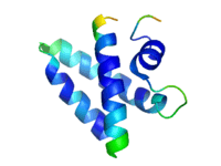
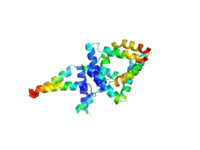
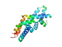
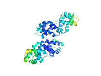
PDB gallery | |
|---|---|
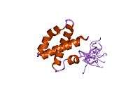 1m12: NMR solution structure of human Saposin C 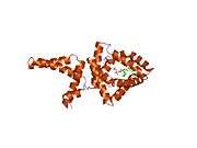 1n69: Crystal structure of human saposin B 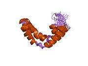 1sn6: NMR solution structure of human Saposin C in SDS micelles 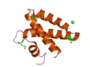 2dob: Crystal Structure of Human Saposin A 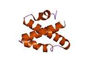 2gtg: Crystal Structure of Human Saposin C |
External links
- Saposins at the US National Library of Medicine Medical Subject Headings (MeSH)