In molecular biology a saposin protein domain is a region of the saposin protein that has a certain conserved sequence or structure that defines a domain. Saposins are small lysosomal proteins that serve as activators of various lysosomal lipid-degrading enzymes.[2] They probably act by isolating the lipid substrate from the membrane surroundings, thus making it more accessible to the soluble degradative enzymes. All mammalian saposins are synthesized as a single precursor molecule (prosaposin) which contains four Saposin-B domains, yielding the active saposins after proteolytic cleavage, and two Saposin-A domains that are removed in the activation reaction. The Saposin-B domains also occur in other proteins, many of them active in the lysis of membranes.[3][4]
Domain organization
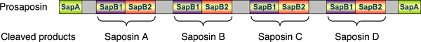
Below is a schematic diagram of the primary structure of the prosaposin protein depicting the N- and C-terminal SapA domains and the four SapB1 and four SapB2 domains. Proteolytic cleavage of the proprotein occurs in the grey regions. Adjacent pairs of SapB1 and SapB2 domains remain connected after proteolytic processing of prosaposin and each pair comprises one of the mature saponin A-D proteins.
Human proteins containing this domain
References
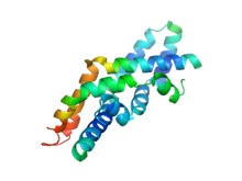
- ↑ PDB: 2qyp, Rossmann M, Schultz-Heienbrok R, Behlke J, Remmel N, Alings C, Sandhoff K, Saenger W, Maier T (May 2008). "Crystal structures of human saposins C andD: implications for lipid recognition and membrane interactions". Structure. 16 (5): 809–17. doi:10.1016/j.str.2008.02.016. PMID 18462685.
- ↑ Munford RS, Sheppard PO, O'Hara PJ (August 1995). "Saposin-like proteins (SAPLIP) carry out diverse functions on a common backbone structure". Journal of Lipid Research. 36 (8): 1653–63. PMID 7595087.
- ↑ Ponting CP (February 1994). "Acid sphingomyelinase possesses a domain homologous to its activator proteins: saposins B and D". Protein Science. 3 (2): 359–61. doi:10.1002/pro.5560030219. PMC 2142785. PMID 8003971.
- ↑ Tschopp J, Hofmann K (March 1996). "Cytotoxic T cells: more weapons for new targets?". Trends in Microbiology. 4 (3): 91–4. doi:10.1016/0966-842X(96)81522-8. PMID 8868085.