Root analogue dental implant
| Root analogue dental implant | |
|---|---|
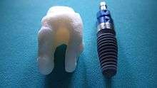
 Multi-rooted ceramic RAI |
A root-analog dental implant (RAI) – also known as a truly anatomic dental implant, or an anatomical/custom implant – is a medical device to replace one or more roots of a single tooth immediately after extraction. In contrast to common 'off the shelf' titanium screw type implants, these implants are custom-made to exactly match the extraction socket of the specific patient.
As the root analog dental implant matches the tooth socket (dental alveolus) it can only be placed in conjunction with the tooth extraction. Thus, if the tooth has been already lost and the soft and hard tissue is already healed a RAI can no longer be placed.
The basic principle of endosseous implants is a biological process described as osseointegration, in which materials such as titanium or ceramic form an intimate bond to bone. There are no particular differences between the osseointegration of a root analog implant and a conventional screw type implant.
The scope of anatomic dental implants is to fit as perfectly as possible into the bony walls of a tooth socket. By adapting the implant to the patient instead of adapting the patient to a rotationally symmetric screw type implant, any surgery on hard or soft tissue, by drilling healthy bone and filling gaps with cadaver/artificial bone, is absolutely unnecessary.
The Problem with conventional implants
As technology has improved, so has implant success rate. However, this does not address fundamental problem with conventional implant technology: the patient must be altered to fit the implant, rather than the other way around.
Screw-type implants are highly unnatural in form and color, and – most importantly – they don’t fit the tooth socket. Even a single-rooted tooth is nearly twice as wide in one direction as in the other. A cylindrical screw implant must be placed into bone, and cannot fit into an existing tooth socket without invasive surgery. Such surgery involves drilling into healthy bone, filling gaps between implant and bone either with bone or bone substitutes, and frequently sinus lift procedures.
Titanium screws are prone to peri-implantitis and plaque accumulation leading to further interventions. Esthetic outcome is highly unpredictable.
Root analogue implants

In contrast to conventional screw, plate, or cylinder implants, RAIs are custom made to perfectly fit the tooth socket immediately after tooth extraction. Therefore every implant is as unique as a patient's fingerprint. As an optimized root-form it is much more than a simple 1:1 replica of a tooth. Since it exactly fills the gap left after the tooth is extracted, surgery is rarely needed. The implant can be produced from a copy of the extracted tooth, an impression of the tooth socket, or from a CT/DVT scan[1]. The advantage of a CT/DVT scan is that the implant can be produced before extraction. With the former methods, it takes one or two days to fabricate an implant.
A root analogue implant is usually fabricated from zirconium dioxide (zirconia), although titanium can be used. Zirconium dioxide is doped with small amounts of yttria, which results in a material with superior thermal, mechanical, and electrical properties, and enhanced fracture toughness - ideal for surgical implants. In addition, zirconia is more esthetic in form and color, with no discoloration visible through gums.[2]
True ‘root-form analogue’ or ‘anatomic’ dental implants have been attempted in the past. Those early attempts failed because of insufficient knowledge of healing of cortical and spongy bone, method, material, tooling, and technology. The principle of Differentiated Osseointegration, in conjunction with suitable material and technology, has enabled the first success in this field[3][4][5].
The Principle of “Differentiated Osseointegration”
Differentiated Osseointegration[6] describes the guided equilibrium of bone-to-implant distance, contact and compression, taking into account spongy or cortical bone, in order to achieve secure osseointegration of individual anatomical dental implants.
The design of the implant surface is crucial in integrating all three possible primary bone-to-implant contact scenarios:
- Contact in the area of the exact root replica, for an immediate start of primary osseointegration without bone trauma;
- Distance at the thin buccal and lingual cortical plates, to safely avoid fracture and pressure resorption of this sensitive bone;
- Compression with macro retentions only in areas of spongy bone to maintain safe primary stability during the entire osseointegration phase.
The combination of all these factors is the most important condition for osseointegration of anatomically shaped dental implants.
Technique
Treatment consists of three simple steps:
- Obtain the 3D form of the tooth to be replaced. This is done either through careful tooth extraction and scanning of the root, taking an impression of the tooth socket, or a pre-op CT/DVT scan. The root analogue implant is produced using modern CAD/CAM technology, based on the Principle of Differentiated Osseointegration;
- Gentle placement of the root analogue implant by simply tapping it in. In general, no surgery is necessary. In particular, no sinus lift or invasive surgery is ever necessary. The implant is placed immediately if it has been produced beforehand from a CT/DVT scan, or the next day if root has to be scanned or an impression of the socket is used;
- A protective splint is fitted to protect the implant during the healing period.
Recovery time is very fast as neither soft nor hard tissue is traumatized. Typically, even the day after implant placement there is no swelling, bruising or pain. After 8–12 weeks healing period, the final crown may be fitted by a family dentist.
Advantages
- Can be placed by any family dentist, requiring no specific surgical skills; there are no guidelines besides indications and contra-indications. The implant is placed with the simplest tools in less than a minute.
- Natural form: a custom milled anatomic implant replicates the natural form and color of a tooth, so it simply fits into the tooth socket. Just like the original tooth, a root analogue implant can have single- and multi-rooted forms.
- Biocompatible: zirconia is metal free and is biocompatible.
- No drilling or surgery, or bone augmentation, is necessary. The patient never needs a sinus lift. There is no additional bone loss, in contrast to a conventional implant where bone must be drilled. No antibiotics are necessary.
- Extremely low risk of peri-implantitis: a conventional implant has a screw winding which is prone to peri-implantitis if it is exposed to the mouth environment. A RAI has none of these problems.
- Immediate: a RAI is gently placed into a tooth socket immediately or the next day after tooth removal. Injury to neighboring roots, nerves or sinus is impossible.
- Esthetic: the all-ceramic structure closely resembles a natural tooth in color. Thus there is no discoloration through the gums, as is commonly seen with titanium implants.
- Widely applicable: RAIs can be used in approximately 30% of cases, as opposed to 5% for conventional implants. The technology is completely open to all common methods of crown reconstruction.
- The consequences in case of implant failure are minimal: the patient's anatomy has not been altered (the tooth socket is unchanged), so there is still the option to switch to a conventional treatment (but not the other way around).
Risks and complications
RAIs are a relatively young technology. Form alterations to the RAI, in conjunction with the pathology of patient, can only be carried out by a practitioner with the requisite knowledge, experience and skills. A failure rate of 10% appears high because of the wider indications for this implant solution; almost all failures occur within the first 4 weeks. After this period, it is rare to have an implant fail.
History
Tooth loss is as old as humanity. Examples from history show that it has always made sense to replace a tooth with an implant that is shaped like a tooth.[7] The earliest known dental implant, discovered in Honduras and dating from 600 AD, is that of a Mayan woman who had several implanted incisors carved from sea shells.[7]
In modern times, a tooth replica implant was reported as early as 1969, but the polymethacrylate tooth analogue was encapsulated by soft tissue rather than osseointegrated.[8]
Lundgren and colleagues used titanium in an experimental model of immediate implant placement with bony integration in 88%[9]. A good fit between implant and bone was considered an important factor for implant success. For this reason, Kohal et al. further refined the approach of root-analogue titanium implants by widening the coronal aspect of the implant to compensate for the lost periodontium and provide a better fit between implant and extraction socket. In several instances implant insertion led to fractures of the thin buccal wall of the alveolar bone. An ensuing clinical study revealed excellent primary stability, but, disappointingly, nearly half the implants failed after 9 months.[10]
Hodosh and colleagues were the first to use custom-made root-analogue implants placed into the extraction socket, reducing bone and soft-tissue trauma. Experimental studies with root-identical titanium implants yielded extremely favourable results with clear evidence of osseointegration and clinical stability. The ensuing clinical trial resulted in 100% primary stability at insertion and 1-month follow up. Due to the high failure rate of 48% over the short time period of 9 months, this particular implant system was not recommended for clinical use.
References
- ↑ Evans, Zachary P.; Renne, Walter G.; Bacro, Thierry R.; Mennito, Anthony S.; Ludlow, Mark E.; Lecholop, Michael K. (2018). "Anatomic Customization of Root-Analog Dental Implants With Cone-Beam CT and CAD/CAM Fabrication: A Cadaver-Based Pilot Evaluation". Journal of Oral Implantology. XLIV (1): 15–25.
- ↑ Bollen, CM (2017). "Zirconia: The Material of Choice in Implant Dentistry? An Update". J Dent Health Oral Disord Ther. 6 (6). doi:10.15406/jdhodt.2017.06.00219.
- ↑ Pirker, W; Kocher, A (2008). "Immediate, non-submerged, root-analogue zirconia implant in single tooth replacement". Int J Oral Maxillofac Surg. 37 (3): 293–5. doi:10.1016/j.ijom.2007.11.008. PMID 18272340.
- ↑ Pirker, W; Kocher, A (2009). "Immediate, non-submerged, root-analogue zirconia implants placed into single-rooted extraction sockets: 2-year follow-up of a clinical study". Int J Oral Maxillofac Surg. 38 (11): 1127–32. doi:10.1016/j.ijom.2009.07.008. PMID 19665354.
- ↑ Pirker, W; Wiedemann, D; Lidauer, A; Kocher, A (2011). "Immediate, single stage, truly anatomic zirconia implant in lower molar replacement: a case report with 2.5 years follow-up". Int J Oral Maxillofac Surg. 40 (2): 212–6. doi:10.1016/j.ijom.2010.08.003. PMID 20833511.
- ↑ Pirker, W; Kocher, A (2009). "True Anatomic Immediate Dental Implant Method: A Clinical Case". International Magazine of Oral Implantology (4): 10–14.
- 1 2 Misch, Carl E (2015). "Chapter 2: Generic Root Form Component Terminology". Dental Implant Prosthetics (2nd ed.). Mosby. pp. 26–45. ISBN 9780323078450.
- ↑ Hodosh, M; Shklar, G; Povar, M (1974). "The porous vitreous carbon/polymethacrylate tooth implant: Preliminary studies". J. Prosthet. Dent. 32 (3): 326–334. doi:10.1016/0022-3913(74)90037-7.
- ↑ Lundgren, D; Rylander, H; Andersson, M; Johansson, M; Albrektsson, T. "Healing-in of root analogue titanium implants placed in extraction sockets. An experimental study in the beagle dog". Clin Oral Implants Res. 3 (3): 136–43. PMID 1290794.
- ↑ Kohal, RJ; Hürzeler, MB; Mota, LF; Klaus, G; Caffesse, RG; JR, Strub. "Custom-made root analogue titanium implants placed into extraction sockets. An experimental study in monkeys". Clin Oral Implants Res. 8 (5): 386–92. PMID 9612143.