RHOT1


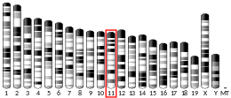
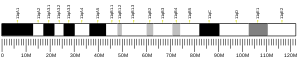
Mitochondrial Rho GTPase 1 (MIRO1) is an enzyme that in humans is encoded by the RHOT1 gene on chromosome 17.[5][6] As a Miro protein isoform, the protein facilitates mitochondrial transport by attaching the mitochondria to the motor/adaptor complex.[7] Through its key role in mitochondrial transport, RHOT1 is involved in mitochondrial homeostasis and apoptosis, as well as Parkinson’s disease (PD) and cancer.[7][8][9]
Structure
In mammals, RHOT1 is one of two Miro isoforms. Both isoforms share a structure consisting of two EF-hand motifs linking two GTP-binding domains and a C-terminal transmembrane domain that attaches the protein to the outer mitochondrial membrane (OMM).[7][10] The EF-hand motifs serve as binding sites for the adaptor protein Milton and the kinesin heavy chain.[11] These domains can also bind calcium ions, and the binding results in a conformational change that dissociates the mitochondrial surface from kinesin.[7][10]
Function
RHOT1 is a member of the Rho GTPase family and one of two isoforms of the protein Miro: RHOT1 (Miro1) and RHOT2 (Miro2).[7][11] Compared to the rest of the Rho GTPase family, the Miro isoforms are considered atypical due to their different regulation.[9] Moreover, the Miro isoforms are only expressed in the mitochondria.[12]
Miro associates with Milton (TRAK1/2) and the motor proteins kinesin and dynein to form the mitochondrial motor/adaptor complex. Miro functions to tether the complex to the mitochondrion while the complex transports the mitochondrion via microtubules within cells.[7][8] Though Miro has been predominantly studied in neurons, the protein has also been observed to participate in the transport of mitochondria in lymphocytes toward inflamed endothelia.[11]
The motor/adaptor complex is regulated by calcium ion levels. At high concentrations, calcium ions arrest mitochondrial transport by binding Miro, causing the complex to detach from the organelle. Considering that physiological factors such as activation of glutamate receptors in dendrites, action potentials in axons, and neuromodulators may elevate calcium ion levels, this regulatory mechanism likely serves to keep mitochondria in such areas to provide calcium ion buffering and active export and, thus, maintain homeostasis.[7]
In addition, Miro regulates mitochondrial fusion and mitophagy in conjunction with mitofusin. According to one model, damaged mitochondria are sequestered from healthy mitochondria by the degradation of Miro and mitofusin. Miro degradation halts their movement while mitofusin degradation prevents them from fusing with healthy mitochondria, thus facilitating their clearance by autophagosomes.[7]
Though the exact mechanisms remain to be elucidated, RHOT1 has been implicated in promoting caspase-dependent apoptosis.[5]
Clinical significance
Studies indicate that Miro may be involved in PD.[8] In neurons, Miro interacts with two key proteins involved in PD, PINK1 and Parkin.[7] Following depolarization of the mitochondria, PINK1 phosphorylates Miro at multiple sites, including S156, and Parkin ubiquitinates Miro, targeting it for proteasomal degradation.[7][8] Degradation of Miro then halts mitochondrial transport.[7]
Though the Rho GTPase family is closely associated with cancer progression, there are few studies demonstrating such association with the atypical Miro proteins. Nonetheless, RHOT1 has been implicated in pancreatic cancer as a tumor suppressor through its regulation of mitochondrial homeostasis and apoptosis. Thus, this protein could serve as a therapeutic target for cancer treatment.[9]
Model organisms
| Characteristic | Phenotype |
|---|---|
| Homozygote viability | Abnormal |
| Recessive lethal study | Normal |
| Fertility | Normal |
| Body weight | Normal |
| Anxiety | Normal |
| Neurological assessment | Normal |
| Grip strength | Normal |
| Hot plate | Normal |
| Dysmorphology | Normal |
| Indirect calorimetry | Normal |
| Glucose tolerance test | Normal |
| Auditory brainstem response | Normal |
| DEXA | Normal |
| Radiography | Normal |
| Body temperature | Normal |
| Eye morphology | Normal |
| Clinical chemistry | Normal |
| Plasma immunoglobulins | Normal |
| Haematology | Normal |
| Peripheral blood lymphocytes | Normal |
| Micronucleus test | Normal |
| Heart weight | Normal |
| Skin Histopathology | Normal |
| Brain histopathology | Normal |
| Salmonella infection | Normal[13] |
| Citrobacter infection | Normal[14] |
| All tests and analysis from[15][16] |
Model organisms have been used in the study of RHOT1 function. A conditional knockout mouse line, called Rhot1tm1a(EUCOMM)Wtsi[17][18] was generated as part of the International Knockout Mouse Consortium program — a high-throughput mutagenesis project to generate and distribute animal models of disease to interested scientists.[19][20][21]
Male and female animals underwent a standardized phenotypic screen to determine the effects of deletion.[15][22] Twenty six tests were carried out on mutant mice and one significant abnormality was observed: no homozygous mutants survived until weaning. The remaining tests were carried out on heterozygous mutant adult mice and no further abnormalities were observed.[15]
Interactions
RHOT1 has been shown to interact with:
References
- 1 2 3 GRCh38: Ensembl release 89: ENSG00000126858 - Ensembl, May 2017
- 1 2 3 GRCm38: Ensembl release 89: ENSMUSG00000017686 - Ensembl, May 2017
- ↑ "Human PubMed Reference:".
- ↑ "Mouse PubMed Reference:".
- 1 2 Fransson A, Ruusala A, Aspenström P (Feb 2003). "Atypical Rho GTPases have roles in mitochondrial homeostasis and apoptosis". The Journal of Biological Chemistry. 278 (8): 6495–502. doi:10.1074/jbc.M208609200. PMID 12482879.
- ↑ "Entrez Gene: RHOT1 ras homolog gene family, member T1".
- 1 2 3 4 5 6 7 8 9 10 11 12 13 14 15 16 17 18 19 20 Schwarz TL (Jun 2013). "Mitochondrial trafficking in neurons". Cold Spring Harbor Perspectives in Biology. 5 (6): a011304. doi:10.1101/cshperspect.a011304. PMC 3660831. PMID 23732472.
- 1 2 3 4 van der Merwe C, Jalali Sefid Dashti Z, Christoffels A, Loos B, Bardien S (May 2015). "Evidence for a common biological pathway linking three Parkinson's disease-causing genes: parkin, PINK1 and DJ-1". The European Journal of Neuroscience. 41 (9): 1113–25. doi:10.1111/ejn.12872. PMID 25761903.
- 1 2 3 Jiang H, He C, Geng S, Sheng H, Shen X, Zhang X, Li H, Zhu S, Chen X, Yang C, Gao H (2012). "RhoT1 and Smad4 are correlated with lymph node metastasis and overall survival in pancreatic cancer". PLOS ONE. 7 (7): e42234. doi:10.1371/journal.pone.0042234. PMC 3409151. PMID 22860091.
- 1 2 Fransson S, Ruusala A, Aspenström P (Jun 2006). "The atypical Rho GTPases Miro-1 and Miro-2 have essential roles in mitochondrial trafficking". Biochemical and Biophysical Research Communications. 344 (2): 500–10. doi:10.1016/j.bbrc.2006.03.163. PMID 16630562.
- 1 2 3 Morlino G, Barreiro O, Baixauli F, Robles-Valero J, González-Granado JM, Villa-Bellosta R, Cuenca J, Sánchez-Sorzano CO, Veiga E, Martín-Cófreces NB, Sánchez-Madrid F (Apr 2014). "Miro-1 links mitochondria and microtubule Dynein motors to control lymphocyte migration and polarity". Molecular and Cellular Biology. 34 (8): 1412–26. doi:10.1128/MCB.01177-13. PMC 3993592. PMID 24492963.
- 1 2 Ogawa F, Malavasi EL, Crummie DK, Eykelenboom JE, Soares DC, Mackie S, Porteous DJ, Millar JK (Feb 2014). "DISC1 complexes with TRAK1 and Miro1 to modulate anterograde axonal mitochondrial trafficking". Human Molecular Genetics. 23 (4): 906–19. doi:10.1093/hmg/ddt485. PMC 3900104. PMID 24092329.
- ↑ "Salmonella infection data for Rhot1". Wellcome Trust Sanger Institute.
- ↑ "Citrobacter infection data for Rhot1". Wellcome Trust Sanger Institute.
- 1 2 3 Gerdin AK (2010). "The Sanger Mouse Genetics Programme: High throughput characterisation of knockout mice". Acta Ophthalmologica. 88: 925–7. doi:10.1111/j.1755-3768.2010.4142.x.
- ↑ Mouse Resources Portal, Wellcome Trust Sanger Institute.
- ↑ "International Knockout Mouse Consortium".
- ↑ "Mouse Genome Informatics".
- ↑ Skarnes WC, Rosen B, West AP, Koutsourakis M, Bushell W, Iyer V, Mujica AO, Thomas M, Harrow J, Cox T, Jackson D, Severin J, Biggs P, Fu J, Nefedov M, de Jong PJ, Stewart AF, Bradley A (Jun 2011). "A conditional knockout resource for the genome-wide study of mouse gene function". Nature. 474 (7351): 337–42. doi:10.1038/nature10163. PMC 3572410. PMID 21677750.
- ↑ Dolgin E (Jun 2011). "Mouse library set to be knockout". Nature. 474 (7351): 262–3. doi:10.1038/474262a. PMID 21677718.
- ↑ Collins FS, Rossant J, Wurst W (Jan 2007). "A mouse for all reasons". Cell. 128 (1): 9–13. doi:10.1016/j.cell.2006.12.018. PMID 17218247.
- ↑ van der Weyden L, White JK, Adams DJ, Logan DW (2011). "The mouse genetics toolkit: revealing function and mechanism". Genome Biology. 12 (6): 224. doi:10.1186/gb-2011-12-6-224. PMC 3218837. PMID 21722353.
Further reading
- Hartley JL, Temple GF, Brasch MA (Nov 2000). "DNA cloning using in vitro site-specific recombination". Genome Research. 10 (11): 1788–95. doi:10.1101/gr.143000. PMC 310948. PMID 11076863.
- Wiemann S, Weil B, Wellenreuther R, Gassenhuber J, Glassl S, Ansorge W, Böcher M, Blöcker H, Bauersachs S, Blum H, Lauber J, Düsterhöft A, Beyer A, Köhrer K, Strack N, Mewes HW, Ottenwälder B, Obermaier B, Tampe J, Heubner D, Wambutt R, Korn B, Klein M, Poustka A (Mar 2001). "Toward a catalog of human genes and proteins: sequencing and analysis of 500 novel complete protein coding human cDNAs". Genome Research. 11 (3): 422–35. doi:10.1101/gr.GR1547R. PMC 311072. PMID 11230166.
- Aspenström P, Fransson A, Saras J (Jan 2004). "Rho GTPases have diverse effects on the organization of the actin filament system". The Biochemical Journal. 377 (Pt 2): 327–37. doi:10.1042/BJ20031041. PMC 1223866. PMID 14521508.
- Brandenberger R, Wei H, Zhang S, Lei S, Murage J, Fisk GJ, Li Y, Xu C, Fang R, Guegler K, Rao MS, Mandalam R, Lebkowski J, Stanton LW (Jun 2004). "Transcriptome characterization elucidates signaling networks that control human ES cell growth and differentiation". Nature Biotechnology. 22 (6): 707–16. doi:10.1038/nbt971. PMID 15146197.
- Wiemann S, Arlt D, Huber W, Wellenreuther R, Schleeger S, Mehrle A, Bechtel S, Sauermann M, Korf U, Pepperkok R, Sültmann H, Poustka A (Oct 2004). "From ORFeome to biology: a functional genomics pipeline". Genome Research. 14 (10B): 2136–44. doi:10.1101/gr.2576704. PMC 528930. PMID 15489336.
- Mehrle A, Rosenfelder H, Schupp I, del Val C, Arlt D, Hahne F, Bechtel S, Simpson J, Hofmann O, Hide W, Glatting KH, Huber W, Pepperkok R, Poustka A, Wiemann S (Jan 2006). "The LIFEdb database in 2006". Nucleic Acids Research. 34 (Database issue): D415–8. doi:10.1093/nar/gkj139. PMC 1347501. PMID 16381901.
- Fransson S, Ruusala A, Aspenström P (Jun 2006). "The atypical Rho GTPases Miro-1 and Miro-2 have essential roles in mitochondrial trafficking". Biochemical and Biophysical Research Communications. 344 (2): 500–10. doi:10.1016/j.bbrc.2006.03.163. PMID 16630562.