Qubit fluorometer
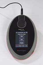
The Qubit fluorometer is a small instrument used for quantification of DNA, RNA, and protein and used in many different applications.[1][2][3][4] The second generation of this instrument, the Qubit 2.0 Fluorometer, was released Nov. 15, 2010. The 3rd generation was launched as Qubit 3.0,and was released in 2014. The current addition to the series is Qubit 4.0 which was launched in the year 2017.
The Qubit fluorometer uses fluorescent dyes to determine the concentration of nucleic acids and proteins in a sample. The UV-absorbance method uses a spectrophotometer to measure the natural absorbance of light at 260 nm (for DNA and RNA) or 280 nm (for proteins). The more DNA, RNA or protein in the sample, the more light is absorbed. The absorbance is a natural property of DNA, RNA, free nucleotides, proteins and some amino acids and many other compounds as well. Because so many molecules absorb light at 260 nm, this measurement is subject to inaccuracy due to potential contamination of the sample with these other molecules. In addition, using the absorbance method, it is not possible to distinguish between DNA, RNA, protein or free nucleotides or amino acids in the sample, leading to potentially highly inaccurate measurements.[5][6][7][8]
The Qubit assays (previously known as Quant-iT) used with both the original Qubit fluorometer and the newer 2.0 fluorometer were developed and manufactured by the previous Molecular Probes (now a part of Life Technologies). Each dye is specific for one type of molecule: DNA, RNA or protein. These dyes have extremely low fluorescence until they bind to their targets (DNA, RNA or protein). Upon binding, they become intensely fluorescent. The difference in fluorescence between bound and unbound dye is several orders of magnitude. For example, the Qubit DNA dye used for the high sensitivity assay has extremely low fluorescence until it binds to DNA. Upon binding to DNA, probably by intercalation between the bases, it assumes a more rigid shape and becomes intensely fluorescent.[9][10] Once added to a solution of DNA, the Qubit DNA dye binds to the DNA within seconds and reaches equilibrium in less than two minutes.
At a specific amount of the dye, the amount of fluorescence signal from this mixture is directly proportional to the concentration of DNA in the solution. The Qubit fluorometer can pick up this fluorescence signal and convert it into a DNA concentration measurement using DNA standards of known concentration. The Qubit fluorometer uses DNA standards to derive the relationship between DNA concentration and fluorescence. It then uses this relationship to calculate the concentration of a sample, based on its fluorescence when mixed with the Qubit dye.
Other fluorometers can also measure the fluorescence from the Qubit dyes and can be used for DNA, RNA and protein quantification in the same way. However, all other fluorometers require the user to use several DNA standards and plot the concentration versus the absorbance on a graph. The data must then be fitted to a line and finally the sample concentration calculated from the equation of the line. Although this is a simple calculation for any scientist, the Qubit fluorometer does this calculation for the user, making it faster and easier, in addition to being less expensive than a typical fluorometer.
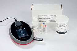
The entire Qubit quantitation system includes the following dyes that are specific for different bio-molecules.
| Reagent/Assay | Assay range | Sample starting concentration range |
|---|---|---|
| Qubit dsDNA HS Assay | 0.2–100 ng | 10 pg/μl–100 ng/μl |
| Qubit dsDNA BR Assay | 2–1,000 ng | 100 pg/μl–1 μg/μl |
| Qubit ssDNA Assay | 1-200 ng | 50 pg/µL-200 ng/µL |
| Qubit RNA Assay | 5–100 ng | 250 pg/μl–100 ng/μl |
| Qubit RNA BR Assay | 20-1,000 ng | 1 ng/µ-1 µg/µL |
| Qubit Protein Assay* | 0.25–5 μg | 12.5 μg/ml–5 mg/ml |
The advantage of using these specific dyes is that the researcher can tell precisely how much of each bio-molecule (DNA, RNA or protein) is in a specific sample, even in the presence of other bio-molecules. For example, one of the DNA quantification kits may be used to measure DNA concentration, one of the RNA kits to measure RNA concentration, and the protein kit to measure protein concentration of the same stock sample. Together, they give the concentrations of DNA, RNA, and protein in a sample.
References
- ↑ Acar E, et al. (2009). "Optimization and validation studies of the MentypeR Argus X-8 kit for paternity cases". Forensic Sci Int: Genet Suppl. 2: 47–48. doi:10.1016/j.fsigss.2009.08.189.
- ↑ Bakos J, et al. (2009). "Enriched environment influences hormonal status and hippocampal brain derived neurotrophic factor in a sex dependent manner". Neuroscience. 164 (2): 788–797. doi:10.1016/j.neuroscience.2009.08.054. PMID 19723563.
- ↑ Halaihel N, et al. (2009). "A new real time PCR-based assay for diagnosing Renibacterium salmoninarum in rainbow trout (Oncorhynchus mykiss) and comparison with other techniques". J Microbiol Meth. 76 (1): 75–80. doi:10.1016/j.mimet.2008.09.014. PMID 18938198.
- ↑ Hamza IA, et al. (2009). "Detection and quantification of human bocavirus in riverwater". J Gen Virol. 90 (Pt 11): 2634–2637. doi:10.1099/vir.0.013557-0. PMID 19656966.
- ↑ Manchester, K.L. (1996). "Use of UV methods for the measurement of protein and nucleic acid concentrations". BioTechniques. 20 (6): 968–970. PMID 8780864.
- ↑ Glasel, J.A. (1995). "Validity of nucleic acid purities monitored by 260 nm/280 nm absorbance ratios". BioTechniques. 18 (1): 62–63. PMID 7702855.
- ↑ Huberman, J.A. (1995). "Importance of measuring nucleic acid absorbance at 240 nm as well as at 260 and 280 nm". BioTechniques. 18 (4): 636. PMID 7598897.
- ↑ Manchester, K.L. (1995). "Value of A260/A280 ratios for measurement of purity of nucleic acids". BioTechniques. 19 (2): 208–210. PMID 8527139.
- ↑ McKnight, R.E., Gleason, A.B., Keyes, J.A., Sahabi, S. (2006). "Binding mode and affinity studies of DNA-binding agents using topoisomerase I DNA unwinding assay". Bioorganic & Medicinal Chemistry Letters. 17 (4): 1013–1017. doi:10.1016/j.bmcl.2006.11.038. PMID 17157016.
- ↑ Schweitzer, C., Scaiano, J.C. (2003). "Selective binding and local photophysics of the fluorescent cyanine dye PicoGreen in double-stranded and single-stranded DNA". Physical Chemistry Chemical Physics. 5: 4911–4917. doi:10.1039/b305921a.