Pyoderma gangrenosum
| Pyoderma gangrenosum | |
|---|---|
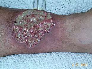 | |
| Pyoderma gangrenosum on the leg of a patient with Crohn's disease. | |
| Specialty |
Dermatology |
Pyoderma gangrenosum is a condition that causes tissue to become necrotic, causing deep ulcers that usually occur on the legs. When they occur, they can lead to chronic wounds. Ulcers usually initially look like small bug bites or papules, and they progress to larger ulcers. Though the wounds rarely lead to death, they can cause pain and scarring.
The disease was identified in 1930. It affects approximately 1 person in 100,000 in the population. Though it can affect people of any age, it mostly affects people in their 40s and 50s.[1]
Types
There are two main types of pyoderma gangrenosum:[1]
- the 'typical' ulcerative form, which occurs in the legs
- an 'atypical' form that is more superficial and occurs in the hands and other parts of the body
Other variations are:[2]
- Peristomal pyoderma gangrenosum comprises 15% of all cases of pyoderma
- Bullous pyoderma gangrenosum
- Pustular pyoderma gangrenosum[3]
- Vegetative pyoderma gangrenosum[4]
Causes
Though the cause is not well understood, the disease is thought to be due to immune system dysfunction, and particularly improper functioning of neutrophils. In support of an immune cause, a variety of immune mediators such as IL-8, IL-1β, IL-6, interferon (IFN)-γ, G-CSF, TNF, matrix metallopeptidase (MMP)-9, MMP-10, and Elafin have all been reported to be elevated in patients with pyoderma gangrenosum.[5]
Also in support of an immune cause is the finding that at least half of all pyoderma gangrenosum patients suffer from immune-mediated diseases.[1] For instance, ulcerative colitis, rheumatoid arthritis, and multiple myeloma (MM) have all been associated with pyoderma gangrenosum. It can also be part of a syndromes such as PAPA syndrome.
One hallmark of pyoderma gangrenosum is pathergy, which is the appearance of new lesions at sites of trauma.[6]
Associations
The following are conditions commonly associated with pyoderma gangrenosum:[7]
- Inflammatory bowel disease:
- Arthritides:
- Hematological disease:
- Autoinflammatory disease:
- Pyogenic sterile arthritis, pyoderma gangrenosum, and acne syndrome (PAPA syndrome)
Diagnosis
Diagnosis of PG is challenging owing to its variable presentation, clinical overlap with other conditions, association with several systemic diseases, and absence of defining histopathologic or laboratory findings. Misdiagnosis and delayed diagnosis are common. It has been shown that up to 39% of patients who initially received a diagnosis of PG have an alternative diagnosis.[9] In light of this, validated diagnostic criteria have recently been developed for ulcerative pyoderma gangrenosum.[10]
Diagnostic Criteria
In addition to a biopsy demonstrating a neutrophilic infiltrate, patients must have at least 4 minor criteria to meet diagnostic criteria.[10] These criteria are based on histology, history, clinical examination and treatment.
- Histology: Exclusion of infection (including histologically indicated stains and tissue cultures)
- Pathergy (ulcer occurring at sites of trauma, with ulcer extending past area of trauma)
- Personal history of inflammatory bowel disease or inflammatory arthritis
- History of papule, pustule, or vesicle that rapidly ulcerated
- Clinical examination (or photographic evidence) of peripheral erythema, undermining border, and tenderness at site of ulceration
- Multiple ulcerations (at least 1 occurring on an anterior lower leg)
- Cribriform or “wrinkled paper” scar(s) at sites of healed ulcers
- Decrease in ulcer size within 1 mo of initiating immunosuppressive medication(s)
Treatment
First-line therapy for disseminated or localized instances of pyoderma gangrenosum is systemic treatment by corticosteroids and ciclosporin. Topical application of clobetasol, mupirocin, and gentamicin alternated with tacrolimus can be effective.
Pyoderma gangrenosum ulcers demonstrate pathergy, that is, a worsening in response to minor trauma or surgical debridement. Significant care should be taken with dressing changes to prevent potentially rapid wound growth. Many patients respond differently to different types of treatment, for example some benefit from a moist environment, so treatment should be carefully evaluated at each stage.
Papules that begin as small "spouts" can be treated with Dakins Solution to prevent infection and wound clusters also benefit from this disinfectant. Wet to dry applications of Dakins can defeat spread of interior infection. Heavy drainage can be offset with Coban dressings. Grafting is not recommended due to tissue necrosis.
If ineffective, alternative therapeutic procedures include systemic treatment with corticosteroids and mycophenolate mofetil; mycophenolate mofetil and ciclosporin; tacrolimus; thalidomide; infliximab; or plasmapheresis.[11]
See also
References
- 1 2 3 Jackson, J Mark; Callen, Jeffrey P (April 23, 2012). Elston, Dirk M, ed. "Pyoderma Gangrenosum". Emedicine.
- ↑ Brooklyn, T.; Dunnill, G; Probert, C (2006). "Diagnosis and treatment of pyoderma gangrenosum". BMJ. 333 (7560): 181–4. doi:10.1136/bmj.333.7560.181. PMC 1513476. PMID 16858047.
- ↑ Shankar, S.; Sterling, J. C.; Rytina, E. (2003). "Pustular pyoderma gangrenosum". Clinical and Experimental Dermatology. 28 (6): 600–3. doi:10.1046/j.1365-2230.2003.01418.x. PMID 14616824.
- ↑ Langan, Sinead M.; Powell, Frank C. (2005). "Vegetative pyoderma gangrenosum: A report of two new cases and a review of the literature". International Journal of Dermatology. 44 (8): 623–9. doi:10.1111/j.1365-4632.2005.02591.x. PMID 16101860.
- ↑ Patel F, Fitzmaurice S, Duong C, He Y, Fergus J, Raychaudhuri SP, Garcia MS, Maverakis E (2015). "Effective strategies for the management of pyoderma gangrenosum: a comprehensive review". Acta Derm Venereol. 95 (5): 525–31. doi:10.2340/00015555-2008. PMID 25387526.
- ↑ Rashid, RM (2008). "Seat belt pyoderma gangrenosum: Minor pressure as a causative factor". Journal of the European Academy of Dermatology and Venereology. 22 (10): 1273–4. doi:10.1111/j.1468-3083.2008.02626.x. PMID 18837131.
- ↑ Brooklyn, Trevor; Giles Dunnill; Chris Probert (2006). "Diagnosis and treatment of pyoderma gangrenosum". British Medical Journal. 333: 181–184. doi:10.1136/bmj.333.7560.181. PMC 1513476. PMID 16858047.
- ↑ Tendas, Andrea; Niscola P; Barbati R; Abruzzese E; Cuppelli L; Giovannini M; Scaramucci L; Fratoni S; Ales M; Neri B; Morino L; Dentamaro T; De Fabritiis P (May 2011). "Tattoo related pyoderma/ectyma gangrenous as presenting feature of relapsed acute myeloid leukaemia: an exceptionally rare observation". Injury. 42 (5): 546–7. doi:10.1016/j.injury.2010.08.014.
- ↑ Weenig, Roger H.; Davis, Mark D. P.; Dahl, Patrick R.; Su, W. P. Daniel (2002-10-31). "Skin ulcers misdiagnosed as pyoderma gangrenosum". The New England Journal of Medicine. 347 (18): 1412–1418. doi:10.1056/NEJMoa013383. ISSN 1533-4406. PMID 12409543.
- 1 2 Maverakis, Emanual; Ma, Chelsea; Shinkai, Kanade; Fiorentino, David; Callen, Jeffrey P.; Wollina, Uwe; Marzano, Angelo Valerio; Wallach, Daniel; Kim, Kyoungmi. "Diagnostic Criteria of Ulcerative Pyoderma Gangrenosum". JAMA Dermatology. doi:10.1001/jamadermatol.2017.5980.
- ↑ Reichrath, Jörg; Bens, Guido; Bonowitz, Anette; Tilgen, Wolfgang (2005). "Treatment recommendations for pyoderma gangrenosum: An evidence-based review of the literature based on more than 350 patients". Journal of the American Academy of Dermatology. 53 (2): 273–83. doi:10.1016/j.jaad.2004.10.006. PMID 16021123.
External links
| Classification | |
|---|---|
| External resources |
| Wikimedia Commons has media related to Pyoderma gangrenosum. |